Acute Myeloid Leukaemia (AML)
Written 23rd July 2020 - Last Update 30th August 2026
Acute Myeloid Leukaemia (AML)
See Leukaemias — Overview for the cross-subtype comparison table.
Myeloid cells are precursors to red cells, monocytes, granulocytes and megakaryocytes. In AML, malignant transformation and uncontrolled proliferation of an abnormally differentiated myeloid progenitor cell results in high circulating numbers of immature blood cells and replacement of normal marrow by malignant cells.
Demographics: incidence rises with age, and AML is the most common acute leukaemia in adults, with a median age of onset of 68 years. Average lifetime risk about 1 in 200. AML comprises approximately 15% of childhood leukaemias.
AML may also occur as a secondary cancer after chemotherapy or radiotherapy for a different cancer, and secondary AML is difficult to treat with chemotherapy alone.
Pattern: malaise + pallor + oral problems.
Classification
Subtyping is now based mainly on the gene or chromosome changes in the AML cells, per the International Consensus Classification and the WHO classification — not on the older morphological grouping. Morphologic criteria from the previous French-American-British system survive only for the not-otherwise-specified subtypes.
Two subtypes matter disproportionately in general practice:
- Acute promyelocytic leukaemia (APL), t(15;17). Represents 10 to 15% of all cases of AML, striking a younger age group with a median age of 31 years. Patients commonly present with a coagulation disorder such as DIC. Suspected APL is a same-day emergency because of the high early death rate from bleeding.
- Therapy-related myeloid neoplasm, from prior alkylating agents, hydroxyurea or topoisomerase II inhibitors. Most occur 1 to 10 years after the initial therapy.
History
Symptoms of acute myeloid leukaemia may be present for only days to weeks before diagnosis, and are usually non-specific. Most arise from disrupted haematopoiesis.
Symptoms that should be investigated:
- Fatigue, pallor or other symptoms of anaemia.
- Symptoms of serious infection — tachycardia, high fevers, rigors.
- Unresolving or unusual infection or fever.
- Abnormal bleeding or bruising.
- Sore gums or mouth ulcers.
- Unexplained bone pain.
- Unintentional weight loss.
Compared with ALL, gingival hypertrophy, stomatitis and skin infiltration are distinctive of AML, while CNS and bone infiltration are less common. Leukaemic cell infiltration of other organ systems tends to be less common in AML than in ALL.
People with AML may only have mild symptoms, and it is not uncommon for a patient with few or no symptoms to be diagnosed unexpectedly on a blood test done in primary care.
Examination
Gingival hyperplasia:

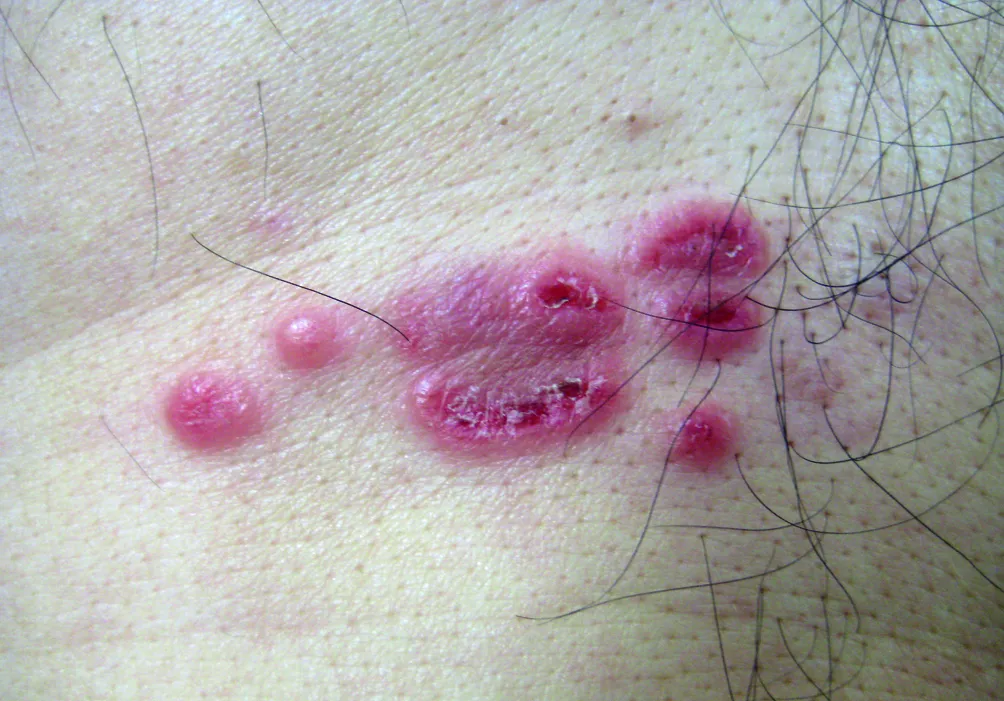
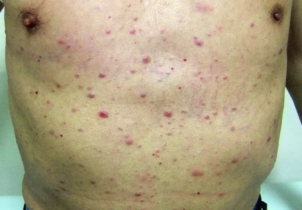
Leukaemia cutis — infiltration of the skin by leukaemic cells. It can have various appearances, including papules or nodules, and plaques, and may be erythematous, brown, haemorrhagic, or violaceous. Usually neither itchy nor painful. Skin biopsy confirms it. Symptoms of extramedullary leukaemic infiltration are present in only approximately 5% of patients, often as skin manifestations.
Investigations in general practice
If a serious blood disorder is suspected, a focused medical history and thorough clinical assessment should be undertaken, and a full blood count and film should be performed immediately.
- FBC and film first — pancytopenia and peripheral blasts suggest acute leukaemia. Blast cells in the peripheral smear may approach 90% of the white cell count.
- Auer rods are pathognomonic for AML, and are not seen in ALL.
- Consider aplastic anaemia, viral infection, and B12 or folate deficiency in the differential of severe pancytopenia.
- A leukaemoid reaction to infection never manifests with high blast counts.
Chase the result the day it is done. Pathology laboratories should directly contact the referring doctor if leukaemia is suspected, and results should be actively followed up by the general practitioner and acted upon on the same day.
Bone marrow aspirate and biopsy is diagnostic; cytogenetics and molecular studies give the prognosis and drive treatment choice.
Referral — this is time-critical
The following signs and symptoms require consultation as a medical emergency:
- Sepsis.
- Symptomatic anaemia.
- Severe thrombocytopenia under 20 × 10⁹/L.
- Major laboratory abnormalities.
- Very high white cell count above 50 × 10⁹/L, or signs of hyperviscosity such as visual disturbance, confusion, severe headache or breathlessness.
- Spontaneous or uncontrolled bleeding.
- Coagulopathy.
Morphologic evidence of APL, disseminated intravascular coagulation, severe thrombocytopenia and any organ dysfunction should be considered a medical emergency.
If the patient is clinically unwell, immediate referral to an emergency facility is recommended without waiting for blood results.
A patient with a laboratory diagnosis of possible AML but no emergency features should be referred for an urgent assessment by a haematologist at an appropriate facility within 24 hours.
Management
Treatment depends on the patient’s overall medical condition, and is best done at the most specialised centre available, particularly during remission induction. Clinical trials are the first choice for most patients where available.
Medically fit patients — induction chemotherapy, then consolidation with or without allogeneic stem cell transplantation. The basic induction regimen (7+3) is cytarabine by continuous IV infusion for 7 days with daunorubicin or idarubicin for 3 days. Complete remission means under 5% marrow blasts, neutrophils above 1 × 10⁹/L, platelets above 100 × 10⁹/L and transfusion independence.
Medically frail or older patients — less intensive therapy. Venetoclax, a BCL-2 inhibitor, is used with azacitidine, decitabine or low-dose cytarabine in adults 75 or older, or with comorbidities precluding intensive induction. Adding venetoclax to azacitidine gave higher remission rates and longer overall survival than azacitidine alone.
Targeted agents by mutation:
- FLT3 — midostaurin added to 7+3, quizartinib for FLT3-ITD, gilteritinib in relapsed/refractory disease.
- IDH1/IDH2 — ivosidenib, olutasidenib, enasidenib.
- CD33-positive — gemtuzumab ozogamicin.
APL is the exception with a good outlook: tretinoin (all-trans retinoic acid) corrects the DIC in 2 to 5 days, and tretinoin with arsenic trioxide, without conventional cytotoxic chemotherapy, is now standard of care in low- and intermediate-risk APL with very high cure rates.
Supportive care the GP shares in
- Febrile neutropenia is an emergency. Febrile patients with neutrophils under 0.5 × 10⁹/L need broad-spectrum bactericidal antibiotics immediately after cultures. Infections progress quickly with little clinical prodrome.
- Transfusion thresholds — prophylactic platelets when the count falls below 10 × 10⁹/L; red cells for haemoglobin below 70–80 g/L.
- Tumour lysis syndrome — hydration plus allopurinol or rasburicase.
- Antifungal and antiviral prophylaxis, and psychological support.
Prognosis
The leukaemia cell karyotype is the strongest predictor of clinical outcome, sorting patients into favourable, intermediate and poor risk groups. Age and comorbidity matter alongside it, and outcomes are generally worse than in ALL.
- Remission induction rate 50 to 85%.
- Long-term (over 5 years) disease-free survival roughly 30 to 40% overall.
- 40 to 50% in younger patients treated with intensive chemotherapy or stem cell transplantation.
Follow-up
Responsibility for follow-up care should be agreed between the lead clinician, the general practitioner, the multidisciplinary team and the patient. Shared follow-up care and specialised care are equivalent in recurrence rate, cancer survival and quality of life.
Usual surveillance schedule in first remission:
- First 2–3 years after treatment: FBC and clinical assessment with a careful history and physical examination every 3 months.
- Thereafter to 5 years: FBC and clinical review every 3 to 6 months.
- Then annual FBC and clinical review indefinitely.
The survivorship care plan should include a process for rapid re-entry to specialist medical services for suspected recurrence, and surveillance for secondary cancers.