Biliary Colic, Cholecystitis & Cholangitis
Written 17th March 2020 - Last Update 27th August 2026
Biliary Colic, Cholecystitis & Cholangitis
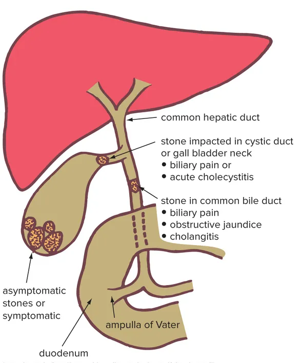
Terminology: choledocholithiasis = gallstones in the common bile duct. Cholecystitis = inflammation of the gallbladder. Cholangitis = inflammation of the bile duct. Gallstones underlie >95% of biliary disease, causing a spectrum from asymptomatic cholelithiasis through biliary colic to acute cholecystitis and acute cholangitis.
Biliary colic
Pain from the biliary tree contracting against an obstructing stone or sludge. Classic demographic female, 40, fat, fair and fertile” — though it can occur in anyone. “Lifetime gallstone risk in first-world countries is 12–20%; ~80% of people with stones stay asymptomatic for life, so cholecystectomy is almost never indicated for asymptomatic stones.
History: Murtagh’s triad — severe pain + vomiting + pain radiation. Epigastric/RUQ pain building to a steady (not truly colicky) crescendo over ~20–30 minutes, lasting at least 30 minutes; radiates to the right shoulder/scapula in ~60%; often nocturnal or ~2 hours after a fatty meal. Pain lasting >6 hours is a red flag for complicated disease (cholecystitis, choledocholithiasis, cholangitis) rather than simple colic.
Examination: may be anxious/restless, or entirely normal. Fever/ rigors point away from simple colic towards cholecystitis or cholangitis. Jaundice suggests a common bile duct (CBD) stone.
Investigations: RUQ ultrasound first-line (>95% sensitive/specific for stones ≥2mm; point-of-care ultrasound performs similarly). LFTs and bilirubin for choledocholithiasis (normal LFTs have ~97% negative predictive value); lipase for pancreatitis. If gallstones are confirmed, ASGE/ESGE risk-stratify for a CBD stone: high risk (cholangitis features, CBD stone seen on ultrasound, or marked cholestasis + duct dilation) → straight to ERCP; intermediate (abnormal LFTs, age >55, or duct dilation alone) → MRCP/endoscopic ultrasound first; low (normal LFTs and ultrasound) → straight to cholecystectomy.
Management: NSAIDs are first-line analgesia (at least as effective as opioids); IV morphine is a reasonable alternative. Keep the patient fasted. No evidence-based role for ursodeoxycholic acid or lithotripsy — not recommended even in patients unfit for surgery. Laparoscopic cholecystectomy is definitive; recent guidelines favour early/index-admission surgery over delayed interval surgery. Choledocholithiasis: ERCP with sphincterotomy and stone extraction, then interval (or same-anaesthetic) cholecystectomy.
Disposition: uncomplicated colic that settles can be discharged with strict return precautions (worsening pain, fever, jaundice) and timely (1–2 week) surgical follow-up for elective cholecystectomy. Watch for complications: acute cholecystitis, obstructive jaundice, cholangitis, gallstone pancreatitis (migrating stones cause up to 50% of cases).
Acute cholecystitis
Persistent cystic duct obstruction, usually by an impacted stone (~90%), causing gallbladder distension, inflammation and secondary infection. Up to 20% of cases develop a complication (empyema, gangrene/ perforation, pericholecystic abscess); diabetics have a higher risk of gangrenous cholecystitis, and the elderly a higher perforation risk if treatment is delayed.
Recognition: steady severe RUQ/epigastric pain and tenderness, worse on deep inspiration; nausea/vomiting in ~75%; positive Murphy’s sign (specificity ~96%, less reliable in the elderly); ± fever, ± jaundice (~15%). No single sign excludes it — diagnosis rests on the combination of local signs, systemic signs (fever, raised CRP/WCC) and supportive imaging (Tokyo Guidelines 2018 criteria). Ultrasound (gallbladder wall >4mm, distension, pericholecystic fluid, sonographic Murphy sign) is first-line imaging; CT or a HIDA scan if ultrasound is equivocal, especially for suspected acalculous disease (below).
Initial management: admit, NPO, IV fluids, analgesia, empiric IV antibiotics, then refer to general surgery.
Empiric IV antibiotics (Therapeutic Guidelines, acute calculous cholecystitis, no sepsis) — kept here for reference:
- 2-drug: gentamicin (or tobramycin) plus amoxicillin 2g IV 6-hourly (or ampicillin 2g IV 6-hourly)
- Non-aminoglycoside alternative (preferred if IV therapy will run ≥72h): ceftriaxone 2g IV daily, or amoxicillin+clavulanate 2+0.2g IV 8-hourly
- Penicillin allergy: non-severe → ceftriaxone; severe immediate → gentamicin/tobramycin alone
- Stop antibiotics immediately after cholecystectomy if no sepsis — no benefit from continuing postoperatively. If septic or surgery is delayed: culture-directed; oral step-down amoxicillin+clavulanate 875+125mg 12-hourly (or trimethoprim+sulfamethoxazole 160+800mg 12-hourly if penicillin-allergic); total IV+oral duration ≤7 days
- Sepsis/septic shock: manage per local biliary-sepsis pathway, not detailed here
Surgery/disposition: early laparoscopic cholecystectomy (within 72 hours) is preferred over delayed surgery — less morbidity, shorter stay, no increased conversion risk. Percutaneous cholecystostomy for acalculous disease or high operative risk (resolves ~90% of cases). The elderly should be treated expeditiously given their higher complication risk.
Acute acalculous cholecystitis — an uncommon variant (~5–10%), without stones, from gallbladder wall ischaemia rather than obstruction; seen in the critically ill, post-operative, diabetic or immunosuppressed. Mortality is much higher (30–90% in critically ill patients). HIDA scan is most reliable when ultrasound is equivocal; early cholecystostomy/cholecystectomy is essential. Therapeutic Guidelines empiric therapy (broader, to cover Pseudomonas/anaerobes): gentamicin/tobramycin plus metronidazole 500mg IV 12-hourly plus amoxicillin 2g IV 6-hourly, or piperacillin+tazobactam 4+0.5g IV 6-hourly alone; total duration just 4 days after source control.
Acute cholangitis
A biliary emergency needing both infection and obstruction — usually gallstones (65%) or a malignant/benign stricture. E. coli, Klebsiella and Enterococcus predominate. Mortality is <10% after drainage but approaches 100% if untreated — early antibiotics ± urgent decompression are critical.
Pitfall: Charcot’s triad (pain, fever, jaundice) is widely taught but has poor sensitivity (26–42%), and Reynolds’ pentad (plus altered mental status and shock) even worse (~5%) — their absence does not exclude cholangitis.
Recognition/diagnosis: the Tokyo Guidelines 2018 criteria (systemic inflammation + cholestasis + imaging evidence of obstruction) are the validated standard. Take blood cultures (up to 70% positive) before antibiotics. Ultrasound first per Tokyo Guidelines (convenience/cost), though CT is increasingly used first-line as it better identifies the obstructing cause and complications; MRCP if no stone is seen.
Empiric IV antibiotics (Therapeutic Guidelines, no sepsis) — kept here for reference:
- 2-drug: gentamicin (or tobramycin) plus amoxicillin 2g IV 6-hourly (or ampicillin 2g IV 6-hourly); add metronidazole 500mg IV 12-hourly if there is chronic biliary obstruction
- Non-aminoglycoside alternative: ceftriaxone 2g IV daily (or cefotaxime 2g IV 8-hourly) — add metronidazole if chronic obstruction — or piperacillin+tazobactam 4+0.5g IV 8-hourly alone (metronidazole not needed with this option)
- Penicillin allergy: non-severe → ceftriaxone/cefotaxime; severe → seek expert advice
- Oral step-down: amoxicillin+clavulanate 875+125mg PO 12-hourly (8- hourly if no biliary drainage performed), or trimethoprim+ sulfamethoxazole 160+800mg PO 12-hourly (plus metronidazole 400mg PO 12-hourly if chronic obstruction) if penicillin-allergic
- Total duration: 7 days if no biliary drainage performed; 4 days after drainage if drainage was performed
- Sepsis/septic shock: manage per local biliary-sepsis pathway, not detailed here
Disposition: ~80% respond to antibiotics alone; the rest need biliary drainage. Endoscopic (ERCP) drainage within 24 hours is first-line, especially with septic shock; transfer early if the local hospital can’t provide endoscopic/percutaneous drainage or ICU care. Early (same-admission) cholecystectomy once infection resolves is safe and may be preferred over delayed surgery.
Gallbladder polyps
Found incidentally on ~0.5% of abdominal ultrasounds; most (65–97%) are benign cholesterol polyps, but malignancy risk rises with size.
Management by size: ≤5mm — discharge, no follow-up; 6–9mm — surveillance (2–5 yearly); ≥10mm — refer for cholecystectomy (malignancy risk 7.6–8.5%). Also refer regardless of size if: age >50, primary sclerosing cholangitis, North Indian/East Asian/Indigenous American ethnicity, growth ≥2mm within 24 months, or symptomatic.
Biliary tract tumours
Gallbladder cancer (mostly adenocarcinoma) and cholangiocarcinoma both often mimic biliary colic/cholecystitis or present late — painless jaundice is the classic presentation of perihilar cholangiocarcinoma (50% of cases). Primary sclerosing cholangitis is the main Western risk factor. Most cases are diagnosed at an advanced, largely palliative stage; surgery is the only potentially curative option, so unexplained/ atypical biliary symptoms in an older patient warrant a lower threshold for imaging and referral.
Care after cholecystectomy
Persistent/recurrent symptoms (“post-cholecystectomy syndrome” — nausea, bloating, jaundice, diarrhoea, pain) occur in up to 40% of patients. Choledocholithiasis (retained stone, ~10–15%) is the most common identifiable biliary cause — work up in general practice or via referral with LFTs (incl. bilirubin) and a RUQ ultrasound, escalating to MRCP if LFTs are abnormal or the CBD is dilated. Bile duct injury (0.1–0.5%) is rarer but the most serious cause — presents early (days post-op) with malaise, fever, jaundice and RUQ pain; refer urgently back to the surgical team.
Prevention
Maintain a healthy body weight (gradual, not rapid, weight loss — rapid loss itself raises stone risk); good glycaemic control; regular moderate activity; a regular eating pattern supports gallbladder emptying. No evidence-based role for pharmacologic prevention.