Cholesteatoma
Written 2nd July 2020 - Last Update 22nd July 2026
Cholesteatoma
Condition
A benign cystic lesion arising from abnormal growth of keratinising squamous epithelium in the soft tissue and bone of the temporal bone — if untreated, can progressively expand and destroy surrounding structures through necrosis and secondary infection.
Demographics: most cases are acquired, usually due to chronic middle ear disease (chronic tympanic membrane retraction is the key mechanism — in a child with hearing loss and chronic retraction, grommet insertion can help prevent progression to cholesteatoma). Congenital cholesteatoma is less common, thought to result from residual embryonal epithelial tissue in the middle ear.
History and examination
Symptoms of eustachian tube dysfunction; history of recurrent ear infections.
Red flags: meningitis-type features; cranial nerve deficits; sensorineural hearing loss; persistent deep ear pain.
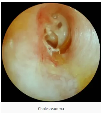
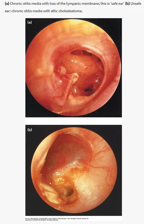
Otoscopy: deep retraction pocket in the attic or posterosuperior quadrant of the tympanic membrane, often with accumulation of squamous debris (the hallmark sign of acquired cholesteatoma); focal granulation on the surface of the drum, particularly at the periphery.
Investigations
Audiometry to assess hearing deficits before planning therapy. Imaging: CT is the modality of choice; MRI in paediatric patients or those with a contraindication to CT.
Management
Refer to ENT for surgical management — generally mastoidectomy.