Vasculitis
Written 2nd April 2020 - Last Update 21st July 2026
Vasculitis
Condition
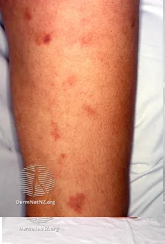
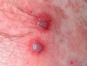
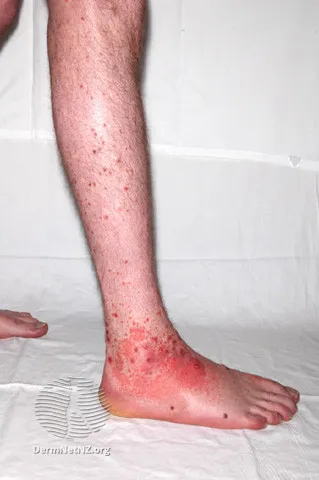
An inflammatory process affecting blood vessel walls; clinical appearance varies with the site, size, and depth of the vessel involved. Palpable purpura is the classical cutaneous lesion, occurring most commonly on the legs.
Three main mechanisms: direct injury to the vessel wall by bacteria/virus, indirect injury by activation of antibodies, and indirect injury through activation of the complement pathway.
Broadly classified by vessel size:
- Large vessel — giant cell arteritis, Takayasu arteritis.
- Medium vessel — polyarteritis nodosa (middle-aged), Kawasaki disease (children).
- Small vessel — leukocytoclastic vasculitis, urticarial vasculitis, chilblains, IgA vasculitis/Henoch-Schönlein purpura (children), acute haemorrhagic oedema (infants).
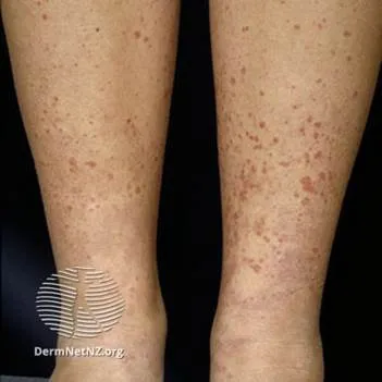
- Capillaritis — progressive pigmented purpura.
Clinical features
Presentation depends on vessel size:
- Capillaritis — pigmented purpura, characterised by petechiae resolving with haemosiderin deposition.
- Small vessel vasculitis — palpable purpura. Palpable vs non-palpable purpura is the key clinical differentiator — palpable purpura points to vasculitis, whereas non-palpable purpura suggests a coagulopathy, thrombocytopenia, or meningococcaemia. Leukocytoclastic vasculitis is the most common type: ~50% idiopathic, 15–20% infection-related, 15–20% connective-tissue-disease-related, 10–15% drug-related — not limited to antibiotics; common drugs such as amlodipine (a widely prescribed antihypertensive) can also trigger it, so take a full medication timeline even for “unlikely” culprit drugs.
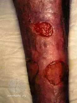
- Medium vessel vasculitis — associated with nodules and livedo reticularis. Polyarteritis nodosa’s systemic features include fever, myalgia, arthralgia, weight loss, and anorexia; the most commonly affected sites are skin, nerves, GI tract, and kidneys, causing pain and neuropathy.
- Large vessel vasculitis — infrequently produces cutaneous features.
Diagnosis is usually made on appearance alone, without further testing; screening tests are done to identify the underlying cause and determine internal organ involvement.
Investigations and complications
HSP needs renal monitoring for nephritis — early morning urinalysis and blood pressure monitoring; renal biopsy may be warranted in severe disease to consider immunomodulator therapy. Polyarteritis nodosa is diagnosed by biopsy of an affected area.
Management
Treatment depends on disease severity, and may include general symptom control, systemic corticosteroids, and immune-modulating agents.
- HSP — largely symptomatic: analgesia (avoid NSAIDs in renal impairment or if using steroids); oral corticosteroid (prednisolone 1–2 mg/kg up to 60 mg daily) if used for more than 5–7 days, taper the dose.
- Leukocytoclastic vasculitis — usually self-limiting, symptoms lasting ~3 weeks. In severe cases with ongoing new lesions, refer and consider prednisolone 25–50 mg daily, tapering once new lesions stop appearing.
- Polyarteritis nodosa — symptomatic pain control plus high-dose corticosteroid therapy ± cyclophosphamide/rituximab; prednisolone 60 mg orally daily can be started if IV methylprednisolone is not immediately available.
Henoch-Schönlein purpura (IgA vasculitis)
The most common vasculitis in children, though it can occur at any age.
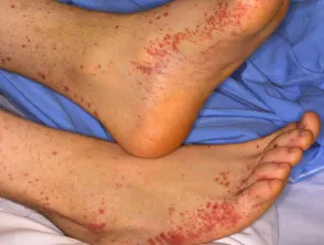
Clinical features: palpable purpura over the lower limbs up to the buttocks; large-joint arthritis; abdominal pain. Typically follows a URTI, associated with streptococcal throat infection (a post-strep complication). Most people recover within a few months.
Complications: IgA nephritis; GI haemorrhage (melaena); intussusception; scrotal involvement.