Squamous Cell Carcinoma (incl. Bowen Disease)
Written 11th March 2020 - Last Update 15th August 2026
Squamous Cell Carcinoma (incl. Bowen Disease)
Condition
Cutaneous SCC is the second most common skin cancer — approximately 28% of all skin cancers — derived from keratin-producing squamous cells of the epidermis. Solar radiation is the major cause; fair-skinned people and those >50 are at higher risk. May be invasive and can sometimes metastasise. Personal history of actinic keratosis is a risk factor; immunosuppression strongly predisposes to SCC. Common on the hands, forearms, head and neck.
Clinical presentations
- Classic SCC — tenderness, thickening, and induration of the lesion; induration/thickening/tenderness in the erythematous base of a scaling lesion strongly suggests early SCC. Hard nodules can form and ulcerate.
- Keratoacanthoma — a rapidly growing lesion with a smooth dome and central keratin core (see Keratoacanthoma for the standalone management approach).
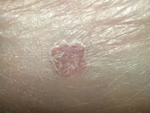
- SCC in situ (Bowen disease / intraepidermal SCC) — a slowly expanding, well-demarcated, erythematous, scaling patch, often mistaken for treatment-resistant psoriasis or dermatitis; occurs mainly on sun-exposed areas, especially the lower leg. Genital and subungual lesions correlate strongly with HPV.
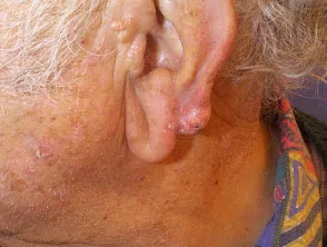
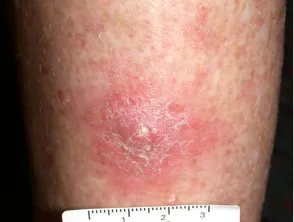
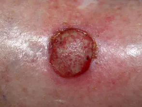
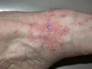
High-risk features
Correlate with higher rates of positive margins, locoregional recurrence, and surgical complications: size >20 mm on trunk/extremities or >10 mm on cheek/forehead/scalp; rapid growth; recurrent/previously incompletely excised lesions; fixation to underlying structures; proximity to significant nerves; poorly defined borders; nodal metastasis; high-risk sites (central face, nose, ears, lips, periocular, scalp, digits, genitalia, hands, below the knee); immunosuppression. High-risk histological features: depth >6 mm, perineural/lymphovascular invasion, extension beyond subcutaneous fat, moderate-to-poor differentiation, or spindle cell/acantholytic/adenosquamous variants. If SCC is suspected, examine regional lymph nodes — regional disease typically develops within 12–24 months of the primary lesion.
Investigations
Biopsy (punch or shave) to confirm the diagnosis; lesions initially thought to be actinic keratosis that persist after cryotherapy, enlarge, or become tender should also be biopsied. If lymph node metastasis is suspected: imaging (CT/ultrasound) of the relevant nodal basin plus fine-needle biopsy.
Management
Surgical excision is first-line. Well-defined, favourable-histology SCC <2 cm can be excised with a ≥4 mm margin (a very high chance of complete excision). Larger or high-risk-site lesions may need up to 10 mm margins — consider specialist referral (plastic surgeon, dermatologist, general surgeon, or GP with advanced excision skills), particularly for central face, scalp, lip and ear lesions given their 10–20% recurrence/ nodal-metastasis rate. A histological margin ≤1 mm mandates consideration of further therapy; incompletely excised SCC has a recurrence rate of ≥50% and should be prophylactically re-excised or treated with radiotherapy.
Bowen disease (SCC in situ) — consider photodynamic-therapy referral, especially in poor-healing sites (e.g. lower leg in older patients). Options: fluorouracil 5% cream daily for 3–4 weeks (repeat once if not fully resolved); excision for smaller well-demarcated lesions; cryotherapy (a single 30-second freeze, 3 mm margin — very effective, but topical therapy is not appropriate if disease extends down hair follicles).
Radiotherapy should be considered for high-risk disease: T4 tumours, rapid growth, recurrence, close margins (<5 mm), perineural/lymphovascular invasion, in-transit metastases, or regional nodal involvement — or where the patient is elderly/frail, the lesion is very large/awkward, or the patient refuses surgery.
Recurrence and follow-up
Within 3 years of a first BCC or SCC, 44% develop a second BCC and 18% a second SCC. SCC recurrence depends heavily on size, site, and histology; high-risk features (>2 cm, lip/ear location, poor differentiation, >4 mm thick, recurrent SCC, perineural invasion) carry up to a 50% risk of regional recurrence — perineural invasion is the single most serious risk factor. After treatment: annual skin checks for life; follow-up at 3 months then 6–12-monthly for up to 3 years (3-monthly then 6-monthly if poorly differentiated, lip SCC, or immunosuppressed), including relevant lymph node examination.
Referral
As above for high-risk clinical/histological features, or if excision is beyond general-practice scope; consider Mohs micrographic surgery for large, ill-defined, recurrent, or cosmetically significant facial lesions.