Pityriasis Versicolor
Written 2nd March 2020 - Last Update 6th September 2026
Pityriasis Versicolor
Condition
A superficial yeast infection of the skin (usually the trunk) caused by Malassezia yeasts. “Versicolor” means variable colours — there are two distinct presentations: reddish-brown, slightly scaly patches on the upper trunk, or a hypopigmented area that will not tan, especially on sun-tanned skin. Mainly affects young and middle-aged adults.
Clinical features
Patches of hyperpigmentation or hypopigmentation — well-demarcated pale or tan macules with fine scales, usually limited to the upper trunk (brown on pale skin, white on tanned skin). Sometimes the whole trunk, upper arms, and neck are affected (but not the face). Patches may coalesce; commonly asymptomatic, but may be slightly itchy. Slight scaling when scratched indicates active infection. Scales removed by scraping show characteristic short, stunted hyphae with spores on microscopy. Often recurrent, especially in summer.
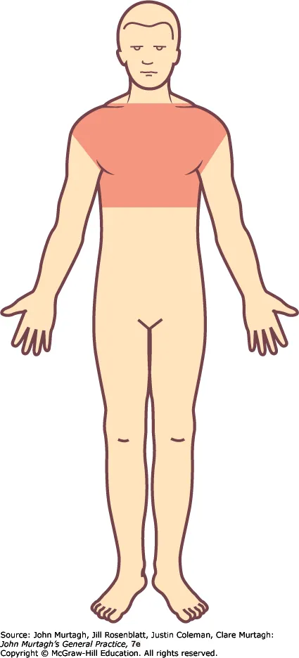
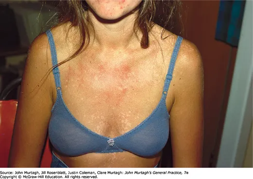
Presentation in skin of colour: Both hypopigmented and hyperpigmented variants occur. In patients with skin of colour, hyperpigmented lesions tend to appear dark brown to grey–black, whereas they are more often light tan or salmon-coloured in patients with lighter skin. Post-inflammatory pigmentary change may persist after clearance of infection and can be a significant source of concern in patients with skin of colour.
Differential diagnosis:
- Seborrhoeic dermatitis of the trunk (more erythematous)
- Pityriasis rosea
- Vitiligo (well-demarcated depigmented macules, no scale)
- Pityriasis alba (hypopigmented with fine scale, commonly on the face and upper limbs of children)
- Confluent and reticulated papillomatosis (reticulated hyperpigmented scaly patches, usually trunk and neck)
- Patch-stage mycosis fungoides (erythematous patches with fine ‘cigarette paper’ wrinkling, bathing suit distribution)
- Erythrasma (well-demarcated hyperpigmented intertriginous patches, showing coral pink fluorescence under Wood’s lamp)
Diagnosis
Usually a clinical diagnosis: characteristic macules and patches with fine scale over the trunk and proximal limbs. Gentle stretching of the affected skin might accentuate the fine scale. The evoked scale sign is a useful bedside finding that is diagnostic of PV. Wood’s lamp examination may show yellow-orange fluorescence.
Confirm with direct microscopy of skin scrapings prepared in potassium hydroxide (KOH), showing hyphae and spores in the characteristic ‘spaghetti and meatballs’ appearance. This simple and minimally invasive test is preferable to a skin biopsy, which might leave scars, particularly in patients with skin of colour.
Treatment
- Econazole 1% topical solution to wet skin, leave overnight, for 3 nights; or
- Ketoconazole 2% shampoo, once daily for 3–5 minutes then wash off, for 5 days; or
- Miconazole 2% shampoo once daily for 10 minutes then wash off, for 10 days; or
- Selenium sulphide 2.5% shampoo, once daily to wet skin, leave on for at least 10 minutes or overnight, for 7–10 days.
In unresponsive cases, oral therapy: fluconazole 400mg orally as a single dose.
Pityriasis versicolor often recurs — repeated courses of topical or oral treatment may be needed. An alternative is prophylactic therapy: itraconazole 200mg twice daily for 1 day per month, for 6 months.
Relapse rates are reported as high as 80%, so maintenance therapy is important. Intermittent use of antifungal shampoos or soaps containing selenium sulphide, zinc pyrithione or ketoconazole is the preventive regimen, e.g. ketoconazole 2% shampoo twice weekly.
Reassure the patient that PV is not contagious, and optimise hygiene such as showering after exercise and washing clothes regularly.
Notes: griseofulvin and terbinafine are ineffective against Malassezia yeasts. Counsel patients that the endpoint of treatment is resolution of scale rather than disappearance of pigmentary change, as pigmentary alteration may persist for many months after successful treatment, particularly in patients with skin of colour. In some dark-skinned patients, pityriasis versicolor causes hypopigmentation that can persist for several months after mycological cure, until sun exposure repigments the pale areas.