Necrotising Fasciitis
Written 2nd March 2020 - Last Update 21st July 2026
Necrotising Fasciitis
See Cellulitis & Skin/Soft-Tissue Infection for the red flags that separate this from ordinary cellulitis.
Condition
A very serious bacterial infection of the soft tissue and fascia (the sheath of tissue covering muscle). Bacteria multiply and release toxins and enzymes that cause thrombosis in blood vessels, resulting in destruction of soft tissue and fascia.
Main types:
- Type I — polymicrobial (more than one bacterium involved). Usually seen in the elderly or in patients with diabetes or other conditions; causative organisms include Staphylococcus aureus, Haemophilus, Vibrio, and several other aerobic/anaerobic strains (Escherichia coli, Bacteroides fragilis).
- Type II — due to haemolytic Group A streptococcus, staphylococci (including MRSA). Sensationalised in the media as “flesh-eating disease”; affects all age groups, including previously healthy people.
- Type III — gas gangrene, caused by Clostridium perfringens or, less commonly, Clostridium septicum. Usually follows significant injury or surgery, and results in gas under the skin causing a crackling sound (crepitus). IV drug users injecting “black tar” heroin subcutaneously can also become infected with clostridia.
- Other — marine organisms (Vibrio species, Aeromonas hydrophila — considered Type III in some reports), usually from wound contamination by sea water, cuts by fish fins/stingers, or consumption of raw seafood; more common with liver disorders, can be fatal if not treated within 48 hours. Fungal necrotising fasciitis (Candida and zygomycetes — Type IV in some reports) complicates traumatic wounds in immunocompromised people.
Necrotising fasciitis affecting the perineal, genital, and perianal regions is known as Fournier gangrene, with a particularly high death rate (15–50%).
Aetiology
An opening in the skin allows bacteria to enter — penetrating/crush injuries, post-surgical procedures. Life-threatening S. pyogenes necrotising fasciitis can occur spontaneously or follow varicella (chickenpox) infection.
Risk factors: aspirin and NSAIDs; advanced age; diabetes; immune suppression; obesity; drug abuse; severe chronic illness; malignancy.
Clinical features
Initial symptoms (within 24 hours of a minor injury): most commonly affects the lower leg, but can also affect the upper limb, perineum, buttocks, trunk, head, and neck. Pain is often severe at presentation and worsens over time. Flu-like symptoms (nausea, fever, diarrhoea, dizziness, malaise) may occur; intense thirst develops as dehydration sets in.
Symptoms after 3–4 days: the affected area swells and may show a purplish rash; large dark marks form, turning into blisters filled with dark fluid; the wound starts to die and blacken (necrosis); oedema is common; a fine crackling sensation under the skin (crepitus) reflects gas in the tissues; severe pain continues until necrosis/gangrene destroys peripheral nerves, at which point the pain subsides (cutaneous anaesthesia). The infection may not improve with antibiotics alone.
By days 4–5: low blood pressure, high temperature, toxic shock; metastatic abscesses can develop in the liver, lung, spleen, brain, pericardium, and rarely the skin.
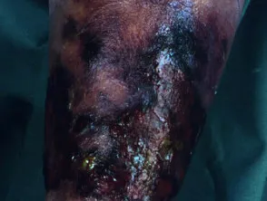
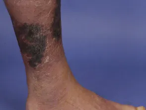
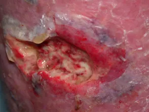
Diagnosis
The finger test is highly pathognomonic: a 2cm vertical incision is made in the affected skin and an index finger is pushed into the tissue — positive if the finger passes through subcutaneous tissue without resistance. Other findings: poor adherence of tissue to fascia on incision; necrotic tissue/pus oozing from fascial planes; dishwater- coloured fluid seeping from the skin. Typically, necrotising fasciitis does not bleed.
Blood tests: high WBC count, low serum sodium, raised CRP, raised CK, raised urea. Blood culture, deep tissue biopsy, and Gram stain help identify the causative organism(s) and guide antibiotic choice. Fungal culture should be performed in immunocompromised and trauma patients. X-ray, CT, and MRI identify fluid collections, inflammation, and gas within the soft tissues.
Management
Surgical removal of devitalised tissue plus urgent antibiotic therapy are essential.
Empirical antibiotics: meropenem 1g IV, or tazocin (piperacillin/ tazobactam) 4/0.5g IV four times daily, plus vancomycin IV, plus clindamycin 600mg IV three times daily (or lincomycin 600mg IV four times daily); add ciprofloxacin 400mg IV three times daily if the wound has been immersed in water.
Confirmed Streptococcus pyogenes: in combination with surgical debridement, a three-drug regimen — benzylpenicillin 2.4g IV 4-hourly (or cefazolin 2g IV four times daily if penicillin hypersensitive), plus clindamycin 600mg IV three times daily (or lincomycin 600mg IV three times daily), plus IVIg as a single dose. Switch to oral amoxicillin 1g three times daily once further debridement is no longer necessary, there’s clinical improvement, and the patient has been afebrile for 48–72 hours.
Confirmed clostridial infection: benzylpenicillin 2.4g IV 4-hourly (or metronidazole 500mg IV three times daily if penicillin hypersensitive), plus clindamycin 600mg IV three times daily (or lincomycin 600mg IV three times daily).
Confirmed MRSA: vancomycin IV, plus clindamycin 600mg IV three times daily (or lincomycin 600mg IV three times daily).
Outcome
If diagnosed and treated early, most patients survive with minimal scarring. Significant tissue loss may need later skin grafting; amputation of limbs may be required to prevent death. Up to 25% of patients die from necrotising fasciitis, due to complications such as renal failure, septicaemia, and multi-organ failure.