Ingrown Toenails (Onychocryptosis)
Written 2nd March 2020 - Last Update 22nd August 2026
Ingrown Toenails (Onychocryptosis)
Condition
A very common condition, especially in adolescent boys, caused by anatomical and environmental factors. Typically located along the lateral edges of the great toenail, representing an imbalance between the soft tissues of the nail fold and the growing nail edge — the basic cause is a redundant skin fold. A skin breach is followed by infection, then oedema and granulation tissue of the nail fold.
Anatomical predispositions (may need surgical correction): congenital malalignment of the nail; hereditary overcurvature of the nail; imbalance between the width of the nail matrix and the nail bed.
Other precipitating factors (usually manageable conservatively): convex or excessive nail cutting; pointed-toe/high-heeled shoes; nail tinea; oral retinoids.
A common problem — accounting for roughly 20% of foot problems seen in primary care. The hallux nail unit is affected in around 80% of cases, and involvement of the lateral nail fold is about twice as frequent as the medial side, likely from repetitive interdigital friction. Onset peaks in adolescence/early adulthood, with a male-to-female ratio of roughly 2:1. Other contributing factors include tight footwear, hyperhidrosis, trauma, digital deformities (hallux valgus, hammertoe), and — for the toenail specifically — oedema from obesity, diabetes, or thyroid/cardiac/renal disease that mimics the soft-tissue crowding of a redundant skin fold.
Staging
Two clinical staging systems are in use, both mapping severity onto the same conservative → antibiotics/surgery → surgical progression this page already follows. The Heifetz classification (the more widely cited):
- Stage I — mild erythema, oedema and pain, without purulence
- Stage II — increased inflammation, with drainage and infection
- Stage III — chronic inflammation with granulation tissue and hypertrophy of the lateral nail fold
DermNet’s equivalent three-stage system ties treatment choice directly to stage: stage 1 managed conservatively (as below); stage 2 may need topical or oral antibiotics, with surgery if it worsens; stage 3 — matching the granulation-tissue/hypertrophy picture above — is usually treated surgically, most successfully with lateral nail avulsion plus matricectomy (the “wedge of nail excision and phenolisation” technique below is one way of performing this).
Initial management on presentation
Should be conservative:
- Gently lift the nail edge and pack moistened cotton wool soaked in 70% alcohol beneath the nail to keep the distal nail plate elevated; repack daily until the toe is healed.
- Remove any nail spicules.
- If bacterial infection: topical antiseptic (e.g. povidone-iodine 10% ointment under occlusion). Oral antibiotics if the lateral nail fold suppurates or cellulitis develops around the toe.
- Treat granulation tissue with curettage and/or light application of a silver nitrate cautery stick.
Prevention
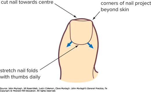
All patients should be instructed on correct foot and nail care: foot baths, avoiding nylon socks, frequent changes of cotton/wool socks. Cotton wool pledgets can be placed beneath the nail edge to assist separation. Cut toenails so the corners project beyond the skin — cut the end of the nail squarely (not the corners) so the nail can grow out from the nail fold; each day after a shower/bath, use the pads of both thumbs to press the nail folds away from the nail.
Treatment — the spiral tape method
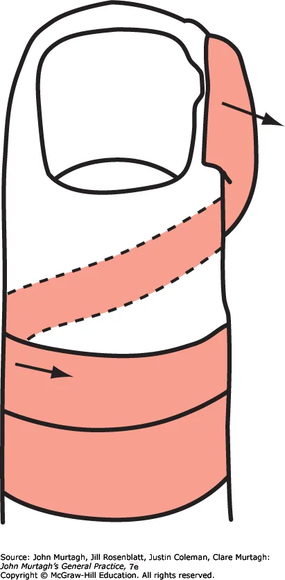
Apply strong adhesive tape (e.g. Elastoplast or Leukosilk 12.5mm) to retract the skin off the ingrowing nail — initially use the thumb pads, despite discomfort, to retract the skin, then pass the tape around the plantar surface to anchor it in loops around the proximal aspect of the toe. Friar’s Balsam on the distal “anchor” gives a better grip. Repeat 2–4 times weekly until the problem settles.
Treatment — surgical
Evidence for surgery over conservative treatment, and for adding phenol. A 2012 Cochrane review (24 RCTs, 2,826 participants) found surgical interventions more effective than non-surgical treatment (gutter treatment/removal, or bracing) at preventing recurrence. Within surgical technique, one trial directly comparing partial nail avulsion with matrix excision, with and without added phenol, found phenol significantly more effective at preventing recurrence (14% vs 41%). The review also looked at postoperative wound care and found no benefit from any of the options tested: prophylactic antibiotics, manuka honey, povidone-iodine with paraffin, hydrogel with paraffin, and plain paraffin gauze all performed the same on infection rate, pain, and healing time — routine postoperative antibiotics for an otherwise uninfected surgical wound aren’t supported by this evidence (distinct from treating an already-suppurating fold before surgery, as above).
- Excision of an ellipse of skin (“army method”) — transposes the skin fold away from the nail; the skin heals, the nail grows normally, and the toe retains its normal anatomy.
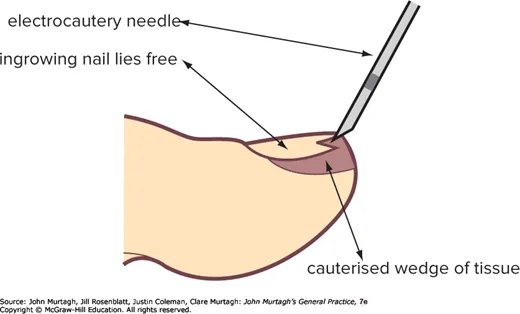
- Electrocautery — similar in principle, but simple, quick and very effective with minimal after-pain, especially for severe ingrowing with much granulation tissue. Under digital block, the electrocautery needle removes a large wedge of skin and granulation tissue so the ingrown nail stands free of skin.
- Skin wedge excision — under digital block, dissect away the skin fold adjacent to the nail, starting from the nail base, extending proximally ~4mm, then sweeping around the side of the nail to under its tip with a 3–4mm margin throughout.
- Wedge of nail excision and phenolisation — excise (with scissors) a wedge of about a quarter of the length of the ingrown nail (rather than a standard wedge resection), then use 80% phenol (concentrated solution) on a cotton wool stick introduced deep into the nail bed space. Warning: take care not to spill phenol onto surrounding skin — it is very corrosive.