Benign & Premalignant Skin Lesions
Written 11th March 2020 - Last Update 28th August 2026
Benign & Premalignant Skin Lesions
Benign lesions
Epidermal (sebaceous) cysts — a firm, skin-coloured, subcutaneous lump, 1–50 mm diameter, tethered to the epidermis but mobile within the dermis; a central punctum with a dark keratin plug may be visible; can become secondarily infected (painful, red). If affecting quality of life or at risk of infection/perforation, excise (minimal excision technique: incise, express contents, remove the cyst wall through a small opening, send for histology to exclude malignancy). Inflamed/infected cysts are difficult to excise — treat infection first (antibiotics ± incision and drainage) and allow inflammation to settle (up to 3–4 weeks) before removal.
Skin tags (fibroepithelial polyps) — painless, skin-coloured, pedunculated growths, 1 mm–5 cm, typically in flexural areas of skin-to-skin friction (axilla, neck, groin).
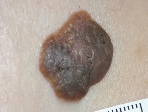
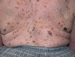
Seborrhoeic keratoses (“senile warts”/“barnacles”) — “stuck-on,” well-demarcated warty plaques, flat or pedunculated, yellow to dark brown, more common after age 40; do not undergo malignant change. Can mimic skin cancer or melanoma but have characteristic dermoscopic features.
For seborrhoeic keratoses and skin tags: reassure if asymptomatic — no treatment is needed. If treatment is wanted, prefer shave biopsy over cryotherapy as it allows histological confirmation; cryotherapy or curettage and cautery are alternatives. These lesions do not generally need excision to exclude malignancy; seek a colleague’s opinion first if in doubt.
Dermoid cysts — a cyst that develops during embryonic skin tissue development, occurring along skin fusion lines; can occur anywhere on the body, but usually the face, neck, or scalp — classically a midline neck lump below the chin. Present in ~40% by birth, most by age 5.
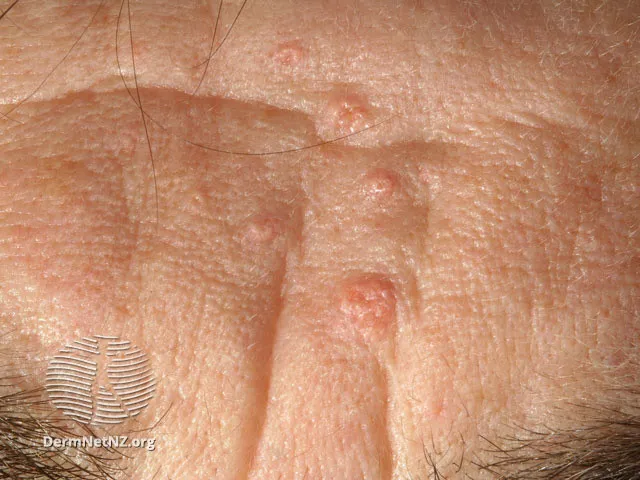
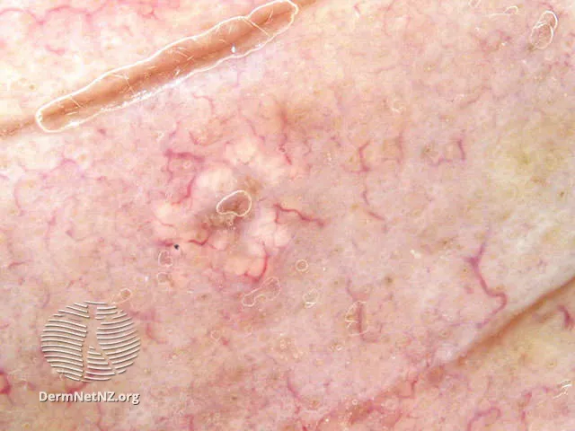
Sebaceous hyperplasia — enlarged sebaceous glands on the forehead or cheeks of middle-aged and elderly patients — can be mistaken for BCC. Small yellow bump up to 3 mm in diameter; close inspection shows a central hair follicle with yellowish lobules and prominent surrounding blood vessels. No treatment required. If bothersome, refer to dermatology for light electrocautery or oral isotretinoin.
Premalignant: actinic (solar) keratosis
Caused by chronic sun exposure; presents as small, red, scaly macules that feel like patches of dry skin, on sun-exposed sites (face, dorsum of hands, forearms, scalp) — can also be atrophic, hypertrophic, pigmented, or lichenoid, and occasionally develop a cutaneous horn. Very common — developed by 40 to 50% of Caucasians aged over 40 years. Considered a precursor lesion to SCC, with annual transformation rates ranging from 0.025% to 20%; in a patient with >10 actinic keratoses, the risk of SCC developing at some stage is 10 to 15%. Thickening or tenderness on lateral palpation suggests possible progression to invasive SCC and should prompt biopsy.
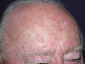
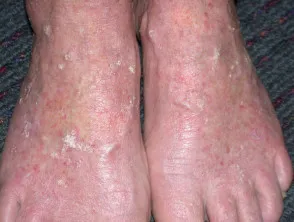
Treatment
It isn’t possible to predict which individual lesions will progress — consider treating all actinic keratoses. Indications: cosmetic appearance, symptom relief, and skin cancer prevention.
- Cryotherapy — cheap and effective. A single ~15-second freeze with a 1 mm margin, healing within ~10 days; longer freezes (for thicker lesions) risk a permanent pale mark/scar. If lesions recur after cryotherapy, consider biopsy to exclude skin cancer.
- Fluorouracil (Efudix) 5% cream — a topical field treatment for widespread/non-clinically-visible changes. Complete clearance in 50% of patients, with a reduction in mean lesion count in 80%. Apply once or twice daily for 2–4 weeks on the face, 3–6 weeks on arms/legs (facial lesions respond faster), until a marked inflammatory response; treated area should not exceed 23 cm × 23 cm at a time. Contraindicated in pregnancy/breastfeeding and DPD deficiency. Common side effects: redness, rawness/discomfort (weeks 2–3), resolving over 2–4 weeks after stopping.
- Imiquimod (Aldara) — second-line. Complete clearance rates of 48 to 57%, partial (>75%) clearance 64–72%. Apply at night, wash off in the morning, usually 3 times a week for 4 weeks (max 2 courses if lesions persist); treated area should not exceed 25 cm × 25 cm. Contraindicated with marked hyperkeratosis, lesions within 1 cm of eyes/nose/lips, immunosuppression (discuss with dermatologist first), or pregnancy/breastfeeding. Side effects can include marked local reactions (up to 4–8 weeks), flu-like symptoms, post-inflammatory hypopigmentation — consider dermatology-supervised first course if extensive reactions are likely.
- Photodynamic therapy — request dermatology referral; also for organ transplant recipients given their higher risk profile.
Dermatology referral is also reasonable if help is needed guiding a patient through their first topical-treatment course.