Basal Cell Carcinoma
Written 11th March 2020 - Last Update 15th August 2026
Basal Cell Carcinoma
Condition
BCC is the most common skin cancer — approximately 60% of all skin cancers. A locally invasive tumour of the basal cells of the epidermis; patients often develop multiple primary tumours over time. Solar radiation is the major cause; usually age >35, more frequent in males. BCC is very rarely life-threatening — a tiny proportion grow rapidly, invade deeply, or metastasise to local nodes.
Clinical presentations
- Nodular — shiny, translucent, telangiectatic papule/nodule; predominates on the head and neck.
- Superficial — bright pink, shiny, well-defined erythematous macule; predilection for the trunk.
- Morphoeic — pale, scar-like, infiltrating plaque, more visible when the skin is stretched.
- Basosquamous — pearly, sometimes with a keratotic component.
Stretching the skin makes all variants more apparent. Ask about change, rapid growth, tenderness, or bleeding; consider skin cancer examination for all patients over 40.
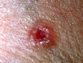
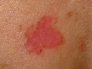
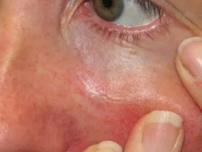
High-risk features
These correlate with a higher likelihood of positive excision margins, locoregional recurrence, and surgical complications: size >20 mm on trunk/ extremities or >10 mm on cheek/forehead/scalp; rapid growth; recurrent or previously incompletely excised lesions; fixation to underlying structures; proximity to significant nerves (e.g. facial, accessory); poorly defined borders; regional lymph node metastasis; high-risk sites (central face, nose, ears, lips, periocular, scalp, digits, genitalia, hands, below the knee); immunosuppression (transplant recipients, CLL, HIV). High-risk histological features: depth >6 mm, perineural/lymphovascular invasion, extension into subcutaneous fat, or micronodular/infiltrative/morphoeic/ basosquamous subtype.
Investigations
Biopsy confirms the diagnosis and subtype.
Management
First-line is surgical excision. Low-risk lesions (well-defined, trunk/extremities <15 mm, face/scalp <10 mm, <6 mm thick, favourable histology) can be excised in general practice with a 2–3 mm margin (commonly 3–4 mm) — adequate microscopic margin is 0.5 mm. Aggressive subtypes (micronodular, infiltrating, morphoeic) need a 4–5 mm margin. Incompletely excised BCCs should be considered for re-excision to achieve clear margins; radiotherapy is a reasonable alternative.
Alternatives when excision is unsuitable/undesired (biopsy-proven superficial BCC only): liquid nitrogen cryotherapy (30-second freeze, thaw, second 30-second freeze, 3–5 mm margin — a longer regimen than for actinic keratosis, and often leaves a permanent pale mark); imiquimod 5% cream (5 times weekly for 6 weeks); curettage and cautery/shave excision (operator-dependent, no histological margin confirmation — use caution on the lower legs of elderly patients); photodynamic therapy (dermatology referral).
Other options: Mohs micrographic surgery for facial BCCs that are large, ill-defined, recurrent, or cosmetically significant; radiotherapy for patients unsuitable for surgery (elderly/frail, very large/awkward lesion, incomplete margins, high-risk histology, or surgery refusal).
Recurrence and follow-up
Within 3 years of a first BCC: 44% will develop a second BCC, 18% a second SCC. 66% of BCC recurrences arise within 3 years of initial treatment; metastasis is rare. After treatment, arrange annual skin checks for life, self-reporting of new/changing lesions, and structured non-surgical-treatment follow-up (3 months, then 6–12 monthly for up to 3 years). Consider closer dermatologist-led monitoring for chronically immunosuppressed patients.
Referral
Refer to a dermatologist, plastic surgeon, general surgeon, or GP with advanced excision skills if excision is beyond general-practice scope, or high-risk clinical/histological features are present.