Haematuria
Written 23rd August 2020 - Last Update 25th August 2026
Haematuria
Haematuria may be macroscopic or microscopic. ~15% of people with macroscopic haematuria have a urological malignancy. Macroscopic haematuria may be visible with as little as 1mL of blood per litre of urine, is usually from the lower urinary tract, but can be the first sign of a renal tumour. Haematuria with clots almost always indicates a lower urinary tract source. Microscopic haematuria is classified as glomerular or non-glomerular.
Only ~30% of patients with microscopic haematuria have an identifiable cause (malignant or benign) — good initial history/examination-based risk stratification avoids over-investigating everyone.
Pseudohaematuria — ruling out red urine that isn’t blood
Red urine caused by something other than blood: foods (beetroot, berries), red confectionery, porphyrins (porphyria), free haemoglobin (haemoglobinuria), or myoglobin (rhabdomyolysis) — urine dipstick/microscopy distinguishes these from true haematuria.
Exercise haematuria is true haematuria (not pseudohaematuria) occurring after heavy exercise — classically running, swimming, or rowing. Investigate if recurrent.
Differential diagnosis by severity
| Serious | Moderate | Minor |
|---|---|---|
| Renal carcinoma, uroepithelial cancer, ureteric stones, prostate cancer, hydronephrosis, tuberculosis, polycystic kidney disease, parenchymal renal disease | Kidney stones, UTI, interstitial cystitis, bladder stones | Asymptomatic, prostatic hyperplasia |
Risk factors for urological malignancy
The greatest risk factors are male sex, smoking history, and increasing age. Also consider: occupational exposure (dyes, benzenes, aromatic amines), cyclophosphamide exposure, chronic UTI history, and prior pelvic irradiation — all worth specifically asking about and flagging in a referral. Starting or using anticoagulants/antiplatelets does not exclude the need for further evaluation — these may unmask, not cause, an underlying pathology. Irritative LUTS (frequency, dysuria, urgency) can reflect UTI or inflammation, but a small, significant proportion of patients with bladder carcinoma in situ present with LUTS alone — be cautious about attributing irritative symptoms to infection without confirming it.
History
- Initial haematuria (suggests urethra/prostate) vs terminal haematuria (suggests bladder)
- New sexual partners (e.g. chlamydia/gonorrhoea)
- Pain suggests infection/inflammation/stone; painless haematuria suggests malignancy
- Recent exercise, menstruation, or sexual activity in the last 24 hours
- Family history of renal calculi
- Anticoagulant or cytotoxic (e.g. cyclophosphamide) therapy
- Recent sore throat (glomerulonephritis) or respiratory infection (IgA nephropathy)
- Overseas travel (malaria, schistosomiasis)
Examination
- Blood pressure, weight, oedema
- Petechiae (vasculitis, clotting disorder)
- Abdominal exam for liver/spleen/kidneys; renal angle/flank tenderness (renal colic)
- Prostate exam in men; DRE + PSA should be considered in men over 50 given prostate cancer can present with haematuria in advanced stages
- Pelvic/speculum exam in women to exclude vaginal/cervical/uterine bleeding sources
- Inspect external genitalia for a local bleeding source (sensitively — patients may not volunteer this)
Investigations
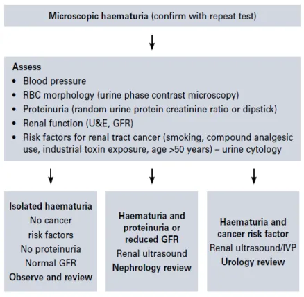
- FBC, UEC, CRP, ESR, coagulation studies, IgA
- Urine dipstick — high false-positive rate; if positive, send formal urinalysis
- Urinalysis + MC&S — if a contaminated specimen is suspected (menstruation, trauma, urogenital atrophy), repeat once resolved; if UTI is found, treat then repeat microscopy 6–12 weeks post-treatment to confirm resolution (or refer on if haematuria persists)
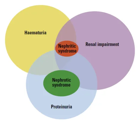
- If proteinuria present: urine protein:creatinine ratio
- Phase-contrast microscopy — dysmorphic red cells suggest glomerular disease
- Urine cytology ×3 — three mid-morning midstream samples on consecutive days; high sensitivity for high-grade tumours/CIS but low sensitivity for low-grade tumours — an adjunct, not a standalone diagnostic tool, particularly useful alongside cystoscopy in patients with upper-tract-cancer/CIS risk factors
- Imaging: renal tract USS is typically sufficient in patients <50 or with significant renal impairment (cheaper, no radiation/contrast risk, but user-dependent and less sensitive for urolithiasis/urothelial lesions). CT IVP (CT KUB with contrast) is the gold standard and preferred in older patients, those with malignancy risk factors, or persistent haematuria
Avoiding unnecessary cystoscopy
Cystoscopy can be avoided if all of: asymptomatic microscopic haematuria, <100 RBC/HPF, normal USS, normal cytology, normal renal function, no proteinuria, no hypertension — these patients often have mild IgA nephropathy and should be monitored 6-monthly (BP, eGFR, MSU, urine protein:creatinine ratio) rather than scoped.
Referral
Urology or nephrology, guided by the suspected bleeding source above. All gross haematuria warrants comprehensive evaluation (urinary tract imaging + urology referral for cystoscopy) to exclude malignancy.
Sources: Murtagh; HNE Community HealthPathways (https://hne.communityhealthpathways.org/17392.htm).