Abdominal Pain
Written 17th March 2020 - Last Update 26th August 2026
Abdominal Pain
Most is benign: acute gastroenteritis and nonspecific abdominal pain are the two single most common diagnoses, followed by cholelithiasis, urolithiasis, diverticulitis, and appendicitis.
Extra-abdominal causes are common too — around 10% of patients over 65 have a respiratory cause.
Examination
Analgesia should be given before a diagnosis is reached. This includes opioid analgesics as they do not increase the risk of diagnostic errors.
Likelihood ratios:
- Murphy sign (cholecystitis): LR+ 15.6. Sonographic Murphy’s sign plus a visible gallstone has 92% positive and 95% negative predictive value.
- Appendicitis: RLQ pain LR+ 7.3–8.5; migratory pain LR+ 3.2; Rovsing sign LR+ 2.5; rebound tenderness LR+ 2 — but over a third of patients with appendicitis have no rebound tenderness, so its absence doesn’t exclude it.
- Diverticulitis: clinical impression, in a patient without complicated-disease risk factors, LR+ 32 — a clinical diagnosis is reasonable without imaging in this group.
- Carnett sign (tenderness increases when a supine patient tenses the abdominal wall by raising legs/shoulders): LR+ 6.5 for abdominal wall pain, LR+ 28 when combined with fixed, localised, superficial tenderness. Useful for chronic abdominal wall pain, but not accurate enough alone to exclude acute intra-abdominal pathology.
- AAA: the classic triad (abdominal/flank pain, hypotension, pulsatile mass) is often incomplete — up to 60% of AAA cases are initially misdiagnosed. Palpation is 68% sensitive/75% specific overall, rising to 100% for aneurysms ≥5cm in a patient with girth <100cm — not useful in a larger patient or smaller aneurysm.
- Testicular torsion: up to 20% of patients have abdominal or flank pain alone — always palpate the testes in male patients with lower abdominal pain.
Investigations
A pregnancy test should be performed in all women of reproductive age with acute abdominal pain, including those using reliable contraception. If positive, pelvic ultrasound follows to locate the pregnancy; keep ectopic pregnancy, adnexal torsion, and PID under consideration.
- CT with IV contrast: first-line for generalised, left-upper-quadrant, or lower abdominal pain when the diagnosis is unclear.
- Ultrasound: first-line for right-upper-quadrant pain (cholecystitis/ cholelithiasis). Point-of-care ultrasound can help with appendicitis, urolithiasis, and AAA at the bedside.
- Pregnancy: ultrasound first-line; MRI preferred over CT if ultrasound is inconclusive.
CT is disproportionately useful in older patients — one large retrospective analysis found it changed the treatment plan more than 65% of the time.
Mesenteric ischaemia (more common in older patients, ~50% embolic, often from atrial fibrillation) needs CT angiography even with renal impairment — a missed or delayed diagnosis is worse than the contrast risk.
Red flags for acute abdominal pain
| History | Signs |
|---|---|
| Collapse at toilet | Pallor and sweating |
| Lightheadedness | Hypotension |
| Ischaemic heart disease | Atrial fibrillation or tachycardia |
| Progressive vomiting, pain, distension | Fever |
| Menstrual abnormalities | Prostration |
| Malignancy | Rebound tenderness and guarding |
| Lack of flatus | Decreased urine output |
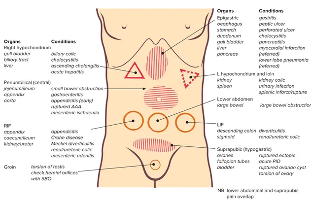
Gynaecological causes of acute lower abdominal pain in women
Consider ectopic pregnancy in all women with new onset low abdominal pain and a positive pregnancy test result.
History: pain characteristics and relation to the menstrual cycle, systemic symptoms, dyspareunia, vaginal bleeding/discharge, contraception, and risk factors for both ovarian torsion and PID.
Examine with speculum and bimanual palpation — cervical motion, uterine, and adnexal tenderness support PID.
Ovarian torsion — a gynaecological emergency:
- Complete or partial rotation of the ovary on its ligamentous supports, obstructing blood supply; prompt diagnosis is needed to preserve ovarian/tubal function.
- Risk factors: reproductive age, a mobile ovarian mass (especially >5cm), prior torsion — though it can occur with normal ovaries too.
- Presentation is often non-specific: sudden-onset pelvic pain, nausea/vomiting, low-grade fever, an adnexal or palpable pelvic mass (not always present); abdominal or pelvic tenderness is present in around two-thirds of patients.
- Pelvic (preferably transvaginal) ultrasound if suspected. Definitive diagnosis requires direct visualisation at surgery.
PID risk factors:
- Age <25
- Recent change of sexual partner
- Current or previous STI, especially repeat infections
- Inconsistent barrier contraception use
- Recent gynaecological procedure (D&C, IVF, IUD insertion, termination)
- Bacterial vaginosis
Clinically unstable (shock/collapse, BP <100/60, pulse >100/min): immediate ambulance transfer. While waiting — large-bore IV access, IV fluids, resuscitate as needed, analgesia.
Clinically stable: refer to the emergency department for suspected appendicitis, peritonism, acute surgical abdomen, ovarian torsion, or ectopic pregnancy with moderate-to-severe symptoms (call the gynaecology registrar first for the last one).
Urgent care and rural/peripheral practice
Elderly patients with abdominal, loin, or flank pain have a 14% mortality rate — symptoms can be vague with low tolerance for shock, so a systolic BP of 90mmHg may already be critical if the patient was previously hypertensive.
In anyone over 50 with abdominal, flank, or back pain: check BP in both arms — a difference of more than 20mmHg is abnormal and can indicate dissection.
A ruptured AAA can mimic renal colic, diverticulitis, or GI haemorrhage.
Complete a testicular examination if the cause of pain is unclear or there’s scrotal pain/swelling; suspected torsion needs urgent escalation, since delayed surgical management risks testicular loss.
Don’t withhold analgesia pending a diagnosis:
- Paracetamol for mild pain
- An opioid for moderate–severe pain
- An antiemetic if nausea/vomiting is present
Send basic pathology to guide the differential:
- FBC/UEC and urinalysis — most presentations
- Urine (and if positive, quantitative serum) βHCG — any woman of childbearing age
- LFTs/lipase — right-upper-quadrant or epigastric pain, or a history of alcohol use
- Troponin — where a cardiac cause is being considered
- INR — if the patient is on warfarin
- Blood cultures — if febrile
Escalate for medical/surgical review per local protocol if pain doesn’t settle with analgesia, or sooner for any red flag above.
Acute abdominal pain in children
Key points: repeated examination is useful to identify persistence or evolution of signs and response to treatment.
Most children need no investigations at all — target them to whatever’s clinically likely.
Appendicitis in young children (under 5) may not present with classic symptoms, and can present as sepsis or perforation, often diagnosed late.
Examination: serial examination of the abdomen is required.
- Peritonism in a child is often positional rather than a fixed sign — the child won’t want to move in bed and can’t walk or hop comfortably; internal rotation of the right hip can irritate an inflamed appendix.
- Genitalia should be examined (e.g. for testicular torsion); a rectal or vaginal exam is rarely indicated in a child and, if needed, should be discussed with a senior clinician and performed only once.
Management: Appropriate analgesia should be used and will not mask potentially serious causes of pain.
Consider consultation with the local paediatric team for:
- A suspected surgical cause
- Severe pain or peritonism
- Bilious vomiting
- Scrotal pain, redness, or swelling
- A child requiring admission
Consider transfer when the required care is beyond the local hospital’s comfort level — ensure adequate analgesia, venous access, and IV fluids first.
Safe for discharge when there are no concerning features, the presentation is consistent with non-specific abdominal pain, a clear follow-up plan is arranged (e.g. with the local GP), and parents have been given advice on when to seek further care.