Palpitations
Written 24th February 2020 - Last Update 26th August 2026
Palpitations
Overview
Palpitations are one of the most common presentations to general practice, and the second most common presentation to cardiologists after chest pain. Most authors describe palpitations as an unpleasant awareness of the heartbeat, sometimes vague and hard for the patient to specify — described as fluttering, skipping, or pounding in the chest or neck, either slow or rapid. Only rarely are they associated with serious underlying disorders or risk of serious cardiac events. In patients presenting to a university medical centre with palpitations: 41% arrhythmic, 31% psychological, 16% unexplained. It is important not to label palpitations as anxiety/panic disorder too readily: 54% of patients given that label go on to be diagnosed with an arrhythmic cause, with a 3.3-year delay to that diagnosis.
Four patterns (EHRA classification) help direct the work-up:
- Extrasystolic — skipped/missed beat, forceful post-ectopic contraction.
- Tachycardic — sudden on/off, fluttering fast heartbeat (gradual onset instead suggests sinus tachycardia).
- Anxiety-related — gradual onset, rate never exceeds what’s physiological for age; often with paraesthesia, hyperventilation, atypical chest pain, globus.
- Pounding — forceful, regular beats at a normal rate; usually a high-output state or structural disease (e.g. aortic regurgitation).
History
Key questions: onset/offset, duration, frequency, triggers, associated symptoms (pre-syncope/syncope, breathlessness, chest pain), existing cardiac conditions.
- Normal heartbeat awareness: forceful beating in the chest that isn’t fast and is regular. Benign; often noticed at night or with anxiety.
- Ectopic beats (atrial/ventricular): often described as skipped beat or sudden forceful contraction in neck. Up to 100 VEBs/24h Holter is normal. There is little evidence that avoiding caffeine reduces ectopy.
- Sinus tachycardia: gradual on/off, usually benign; occasionally thyrotoxicosis or anaemia.
- SVT: sudden on/off, regular, may follow sudden movement (e.g. bending). May have chest discomfort or light-headedness or breathlessness; syncope uncommon. Patients with SVT can frequently terminate an event with vagal manoeuvres such as the Valsalva technique or cold stimulus to the face.
- AF: sustained, sudden-onset, irregular; exertional breathlessness common. When syncope occurs with AF it is more often due to a significant sinus pause at the time of spontaneous reversion (tachycardia-bradycardia syndrome) than the arrhythmia itself.
- VT: rarely isolated palpitations — usually structural heart disease with haemodynamic compromise → emergency transfer.
Discriminating features:
- A systematic review found only six historical factors significantly correlated with an arrhythmic cause: age >60 years, regular palpitations, palpitations affecting sleep, a regular pounding sensation in the neck, visible neck pulsations, and vasovagal symptoms (pallor or diaphoresis).
- Regular neck pounding has a particularly strong positive likelihood ratio (LR+ 177) for AV nodal re-entrant tachycardia when present (LR− 0.07 when absent), though some studies suggest this is less discriminating between arrhythmia types than first thought, since cannon A waves can also occur in ventricular tachycardia and complete heart block.
- Other useful likelihood ratios: a history of cardiac disease (LR+ 2.03), palpitations affecting sleep (LR+ 2.29) or occurring during work (LR+ 2.17) each raise the probability of an arrhythmic cause; duration under 5 minutes lowers it (LR− 0.38).
Ask about predisposing conditions, psychiatric history, and medications/substances. A family history of sudden death in one relative raises the relative risk of sudden death to 1.8-fold, and in two relatives to 9.4-fold — ask over at least three generations. Caffeine intake was not associated with increased arrhythmia risk in a large cohort study even after adjustment for confounders. QT-prolonging drugs (antipsychotics, tricyclics, macrolides, methadone) merit review; taper beta-blockers rather than stopping abruptly. Anxiety and depression are common comorbidities, not alternative diagnoses to be assumed by exclusion: up to 31% of palpitation presentations are ultimately attributed to a psychiatric cause, but up to two-thirds of patients labelled with a psychiatric cause are later found to have a cardiac arrhythmia, most often managed with catheter ablation.
Examination
Examine for cardiac and other underlying systemic illnesses:
- Resting and postural HR/BP.
- Cardiac exam: murmurs, displaced apex (mitral valve prolapse, aortic regurgitation, structural cardiomyopathy).
- Thyroid exam, weight, signs of anaemia.
- Only examination finding significantly associated with arrhythmia = abnormal resting HR (>100 or <60 bpm).
- An irregular pulse has a positive predictive value of 91% for finding a significant arrhythmia on subsequent 24-hour ECG monitoring.
Investigations — rational work-up
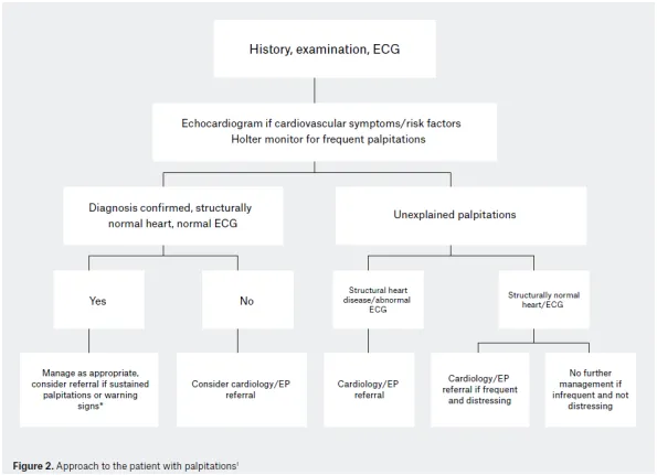
ECG in all patients. Rarely captures symptoms, but the resting sinus-rhythm ECG can still point to a mechanism — see ECG Interpretation for the systematic reading approach:
| Resting ECG finding | Consider |
|---|---|
| Pre-excitation/delta wave | WPW — AVRT |
| Left atrial enlargement, frequent PACs, sinus bradycardia | Atrial fibrillation |
| Left ventricular hypertrophy | Atrial fibrillation, ventricular tachycardia |
| Frequent PVCs | Ventricular tachycardia |
| Q waves | Ischaemic heart disease — AF, VT |
| Widespread T wave inversion, LVH, Q waves and ST changes | Hypertrophic cardiomyopathy — risk of AF, VT |
| Long/short QT, Brugada pattern, early repolarisation | Genetic arrhythmia syndrome — risk of sudden cardiac death |
| Inverted T or epsilon waves in V1–V3 (no RBBB) | ARVC — risk of sudden cardiac death |
Bloods: FBC, UEC, TSH — guided by clinical suspicion, not routine.
Ambulatory ECG monitoring. Match the device to symptom frequency:
| Device | Best for | Diagnostic yield | Trade-off |
|---|---|---|---|
| 24–48h Holter monitor | Daily/near-daily symptoms | ~10–15% | Low yield if less than daily; no patient activation needed |
| Event/loop recorder (1–4 weeks) | Weekly–monthly symptoms | ~50–85% | Mostly patient-triggered; not useful for asymptomatic arrhythmia/syncope |
| Implantable loop recorder | Months–years, sporadic (<1/month) | ~80–90% | High cost; mainly approved for syncope/cryptogenic stroke work-up |
| Handheld ECG (e.g. AliveCor) | Patient wants a permanent option | High (with use) | Cost to patient; activation delay; no Medicare rebate |
Echocardiogram, with symptoms or risk factors for cardiovascular disease:
- Coronary artery disease.
- Family history of sudden cardiac death.
- Abnormal ECG.
- Exertional symptoms.
- Cardiac symptoms -> syncope/chest pain/diaphoresis/shortness of breath.
- Sustained rapid palpitations.
Exercise stress test if symptoms are exertional — provokes and documents the arrhythmia.
Electrophysiology study (EP referral). If clinical suspicion remains high — such as with a classic history of SVT — despite a normal initial work-up, specialist referral should be considered for electrophysiology study with or without ablation or an implantable loop recorder. The advantages of electrophysiology study include identifying the arrhythmia mechanism, potentially curing the arrhythmia during the same procedure, and avoiding the delay required to document arrhythmia on serial ambulatory monitoring — especially pertinent if palpitations are associated with syncope or there is structural heart disease. The risks include the invasive nature of the procedure, including vascular access complications, and the rare risk of damage to the AV node during ablation.
Management and referral
Work-up is frequently normal. If well and without concerning features, reassure.
Which patients to refer, or “when to worry”:
- Patients with frequent or persistent palpitations
- Sustained rapid palpitations
- Significant associated symptoms:
- Pre-syncope/syncope (consider situational context)
- Breathlessness
- Chest pain
- Family history of recurrent syncope or of sudden death
- Significant resting 12-lead electrocardiography or echocardiographic abnormalities
- Wolff–Parkinson–White syndrome (pre-excitation including short PR interval and delta wave)
- Signs of structural or electrical abnormalities:
- T wave abnormalities
- Prior myocardial infarction (Q waves)
- Long or short QT interval, Brugada pattern, early repolarisation pattern
Patients presenting with syncope or ongoing chest pain should be transferred to an emergency department by ambulance rather than managed in general practice. Certain occupations (pilots, commercial drivers) warrant specialist referral for occupational-fitness assessment even for otherwise low-risk findings.
Ectopic beats — detailed management
- Atrial ectopics: usually asymptomatic; reassurance-based. Check lifestyle factors (alcohol, caffeine, stress, smoking). Rarely needs treatment — no ideal anti-ectopic agent. May forerun PSVT or AF. Intolerable symptoms: atenolol 25–100mg daily, metoprolol 25–100mg BD, or verapamil SR 180–480mg daily.
- Ventricular ectopics: usually asymptomatic (90%) — occur in 20% of “normal” hearts. Often felt as “missed beats,” more at rest than exercise. Digoxin and sympathomimetics can cause both types.
- Be careful for: possible forerunner of VT; ventricular dysfunction; ectopic- mediated cardiomyopathy. Rule out ischaemic heart disease, mitral valve prolapse, thyrotoxicosis, LV failure.
- Symptomatic but well, normal CXR/ECG: reassure — a normal echo often helps. Never start drug therapy without an echo first — Class I antiarrhythmics can worsen the arrhythmia or become life-threatening with reduced ventricular function (refer to cardiology if so). Troublesome symptoms: atenolol 25–100mg daily, or metoprolol 25–100mg BD.
- Benign but frequent/symptomatic: atenolol 25mg, metoprolol 25mg BD, or verapamil 180mg.