Infective & Inflammatory Heart Disease
Written 24th February 2020 - Last Update 27th August 2026
Infective & Inflammatory Heart Disease
Pericarditis
Causes: infective (most commonly viral) or non-infective — autoimmune (common), neoplastic, metabolic, traumatic (including post-AMI), iatrogenic.
Diagnosis: pericarditic chest pain is typically sharp and pleuritic, eased sitting up/leaning forward, worsened supine. Inflammatory pericardial syndrome requires at least 2 of 4: pericarditic chest pain; pericardial rub; new widespread ST elevation or PR depression on ECG; new/worsening pericardial effusion. Supporting findings: raised CRP/ESR/WBC, or pericardial inflammation on CT or cardiac MRI (gold standard).
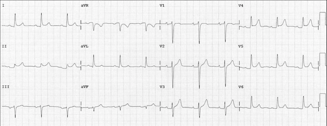
Important considerations: rule out other life-threatening chest pain causes (ACS, PE, myocarditis). Identify clinically significant pericardial effusion, which can progress to cardiac tamponade — classic triad of soft heart sounds, elevated JVP, and hypotension.
Investigations (all suspected patients): ECG, CXR, inflammatory markers, cardiac markers, transthoracic echocardiogram.
Causes and their work-up:
| Cause | Prevalence | Investigation |
|---|---|---|
| Idiopathic | Most common | Diagnosis of exclusion |
| Viral | Most common infectious cause | Serology not routine — diagnosis mainly clinical; consider HIV in high-risk patients |
| Bacterial | Uncommon — can occur with pneumonia | Blood + pericardial fluid cultures |
| Tuberculosis | Uncommon — consider exposure/demographic | CXR, tuberculin testing, QuantiFERON-Gold, sputum culture |
| Fungal/parasitic | Rare in Australia | — |
| Autoimmune | Uncommon — consider with RA, SLE | Target testing to likely disease |
| Neoplastic | Consider with lung/breast/haematological cancer | Pericardial fluid cytology, biopsy |
| Post-procedural | Common after cardiac/thoracic surgery | History |
| Post-MI | Delayed by weeks; more common in large anterior infarcts | — |
| Uraemic | Suspect in CKD, especially peri-dialysis | Urea, creatinine |
| Radiation | Consider with chest irradiation | — |
| Drugs | Rare | — |
Management: admit if a specific treatable cause is found, or with poor-prognosis features:
- High fever >38°C.
- Subacute course.
- Large effusion.
- Tamponade.
- Failure to respond to 7 days of NSAIDs/aspirin.
- Myopericarditis, trauma, immunosuppression.
- Oral anticoagulation (precludes standard treatment).
Treat the specific cause where found (e.g. antibiotics for infective, dialysis for uraemic).
First-line treatment is NSAIDs plus exercise restriction:
- Colchicine 500mcg BD for 3 months (>70kg) or 500mcg daily for 3 months (<70kg or >70yo), plus one of:
- Aspirin 750–1000mg PO 8-hourly for 1–2 weeks, then taper by 250–500mg every 1–2 weeks until stopped, or
- Ibuprofen 600mg PO 8-hourly for 1–2 weeks, then taper by 200–400mg every 1–2 weeks until stopped.
- Use gastric protection with NSAIDs.
Steroids are sometimes used by specialists but aren’t on eTG for acute pericarditis. Previous recurrent-pericarditis regimens used 0.2–0.5mg/kg/day for 2–4 weeks, tapering 10% every 1–2 weeks to 15mg/day, then reducing 1mg per 6 weeks — osteoporosis prophylaxis is needed with protracted corticosteroid use.
Infective endocarditis
Pathogenesis: endocardial injury (usually from turbulent flow) → thrombus formation → seeding by bacteria introduced into the bloodstream → chronic bacteraemia. Organisms: Staphylococcus, then strep viridans, then coagulase-negative staphylococci are the 3 most common; other streptococci and enterococci are also frequent; fungal causes are rare.
High-risk groups: IV drug use, recent dental surgery, elderly, men more than women, structural heart disease.
Suspect IE and apply the modified Duke criteria in patients with: prolonged fever (FUO); fever with vascular phenomena (stroke, limb ischaemia, septic emboli); ≥2 persistently positive blood cultures; a febrile patient with a prosthetic valve, IV drug use, a predisposing heart condition, or recent hospitalisation.
Diagnostic criteria
Major clinical criteria (2 positive = definite): blood cultures positive from 2 separate cultures showing typical organisms, or a single positive culture for Coxiella burnetii or antiphase I IgG antibody titre >1:800; or echocardiographic evidence of endocardial involvement (abscess, new valvular regurgitation — a worsening/changing pre-existing murmur doesn’t count).
Minor clinical criteria: predisposing heart condition or IV drug use; fever; vascular phenomena (major arterial emboli, septic pulmonary infarcts, intracranial or conjunctival haemorrhage, Janeway lesions); immunological phenomena (glomerulonephritis, Osler’s nodes, Roth’s spots, raised rheumatoid factor); microbiological evidence not meeting the major criterion.
Pathological criteria (either = definite): organism demonstrated on culture/ histology of a vegetation; or a pathological lesion (vegetation/intracardiac abscess) showing active endocarditis.
Classification: Definite IE — ≥1 pathological criterion, or 2 major, or 1 major
- 3 minor, or 5 minor. Possible IE — 1 major + 1 minor, or 3 minor. Rejected — consider other causes of fever (other infection, rheumatological, oncological).
Two further routes to a major criterion were added in the 2023 Duke-ISCVID revision: intraoperative findings (vegetations, abscess, valvular destruction, prosthetic valve dehiscence/loosening), used when other definitive criteria are unavailable; and molecular microbiology (amplicon/metagenomic sequencing), increasingly used to identify the organism in blood-culture-negative endocarditis.
Important consideration: spread of infection to other organs and progression of cardiac infection are common — consider particularly in patients failing to improve or deteriorating on antibiotics.
Investigations: transoesophageal echo is more sensitive than transthoracic — cardiac CT and PET/CT are now also accepted imaging modalities alongside echo for major-criterion findings. Take 3 separate blood culture sets before starting antibiotics (identifies ~90% of pathogens).
Management: requires a multidisciplinary team (ID + cardiology + cardiothoracics). Prolonged IV antibiotics — first-line beta-lactam + gentamicin — with directed therapy once sensitivities are known. May require surgery.
Kawasaki disease
An acute self-limiting vasculitis of medium-sized arteries, predominantly affecting children under 5 — the leading cause of acquired heart disease in children in developed countries, via its risk of coronary artery aneurysm formation. The disease runs through three stages: an acute phase (abrupt high fever lasting 1–2 weeks, up to 3–4 if untreated, with rash, conjunctivitis and myocarditis), a subacute phase as the fever subsides through weeks 4–6 (desquamation of the hands, thrombocytosis, and coronary artery aneurysm development), then a convalescent phase with clinical resolution within 3 months, though cardiac abnormalities can persist into this phase and new aneurysms after week 8 are unusual.
Diagnostic criteria: fever for ≥5 days, plus ≥4 of: bilateral non-purulent conjunctival injection; oral mucosal changes (red/cracked lips, strawberry tongue, pharyngeal erythema); polymorphous rash; extremity changes (erythema/oedema acutely, periungual desquamation in the subacute phase); cervical lymphadenopathy (usually unilateral, >1.5cm). Any criterion met at any point during the illness counts toward the diagnosis, even if not present at the time of assessment.
Incomplete Kawasaki disease (fewer criteria met) is increasingly recognised, especially in infants, and still needs echocardiography if suspected given the same coronary artery risk. A CRP/ESR-based algorithm helps decide who needs imaging: with a CRP under 30mg/L and ESR under 40mm/hr, follow daily and get an echocardiogram only if fever persists or desquamation occurs; with CRP over 30 or ESR over 40, get the echocardiogram now. Treat as incomplete KD once more than three supporting features are present alongside the ≥5-day fever. An infant under 6 months with fever beyond 7 days should get an echocardiogram to exclude KD even without meeting the criteria count, since incomplete presentations are more common at the extremes of the paediatric age range.
Investigations: echocardiography at diagnosis, 1–2 weeks, and 4–6 weeks to assess for coronary artery aneurysms; inflammatory markers (CRP/ESR) are typically markedly raised; platelets are often normal or low acutely, rising in the subacute phase — mild to moderate normocytic anaemia is characteristic of the acute phase. Thrombocytopenia (rather than the more typical later thrombocytosis) may occasionally be present in the acute phase and predicts a greater risk of coronary artery aneurysm.
Management: IVIG (2g/kg as a single infusion over 10–12 hours) plus high-dose aspirin (80–100mg/kg/day divided 6-hourly, continued until afebrile for over 48 hours), ideally started within 7–10 days of fever onset, substantially reduces coronary aneurysm risk (from ~25% untreated to ~4%). Aspirin is then continued at a lower antiplatelet dose (3–5mg/kg/day) after the acute phase, for at least 6–8 weeks or longer if aneurysms are present. IVIG-resistant cases (persistent/recurrent fever, roughly 15–20% of patients) — defined as still febrile 36–48 hours after the first infusion — get a second IVIG dose; corticosteroids (extended-course prednisolone, as used in the RAISE trial) are an option particularly in those judged at high risk of IVIG resistance, and infliximab is a further option for refractory disease. All cases need paediatric cardiology involvement and serial echocardiographic follow-up given the aneurysm risk.
Myocarditis
Inflammatory disease of cardiac muscle from a range of infectious and non-infectious causes. Can be acute (developing over ≤3 months) or chronic (>3 months); focal or diffuse; can lead to dilated or other cardiomyopathies. Presentation ranges from subclinical disease to fatigue, chest pain, heart failure, cardiogenic shock, arrhythmias, and sudden death. No population-based epidemiological data exists, since there’s no safe non-invasive diagnostic test and many cases are subclinical/ non-specific.
Diagnosis requires myocardial biopsy for confirmation, though many patients don’t undergo this. Worse outcomes are seen with LVEF <50%, sustained ventricular arrhythmias, or low cardiac output syndrome at presentation. Typical profile: ACS-like symptoms, or new/worsening heart failure (exertional dyspnoea, peripheral oedema, chest discomfort, fatigue over 2 weeks to 3 months), or life-threatening arrhythmias/ cardiogenic shock/severe LV impairment without angiographic evidence of CAD.
Important consideration: myocarditis with associated pericarditis presents with positional/pleuritic pain and pericardial effusion.
Investigations: ECG (ST elevation/depression, T-wave inversion); echo/cardiac MRI (global or regional LV/RV dysfunction); troponin (may or may not be raised); BNP if heart failure is suspected. Cardiac biopsy is the gold standard — biopsy-proven myocarditis carries a worse prognosis.
Management:
- Hospital admission, especially for fulminant myocarditis — new severe heart failure requiring inotropic/mechanical circulatory support. High mortality without transplant: 75% survival at 1 year, 65% at 9 years without transplant, vs 100% at both timepoints with transplant.
- ECG monitoring for both tachy- and bradyarrhythmias, which can be asymptomatic yet life-threatening — a key reason hospitalisation is needed.
- Avoid: NSAIDs (may worsen the inflammatory process), heavy alcohol, and exercise (restrict to reduce cardiac workload).
- Cause-specific therapy: antivirals, immunosuppressants, IVIG, IV antibiotics as indicated.
- Follow-up: cardiology review every 1–3 months; gradual return to physical activity with effort-tolerance monitoring; watch for persistent S3/S4 gallops; regular echo or cardiac MRI/nuclear imaging to reassess function.
- Prevention: vaccination can prevent some forms of viral myocarditis, though its overall preventive role is not well established.
Causes of myocarditis (reference)
Infectious:
- Viral — adenovirus, arbovirus, Coxsackie B, CMV, dengue, echovirus, EBV, hepatitis B/C, herpesvirus, HIV, influenza A/B, mumps, parvovirus, poliomyelitis, rabies, rubella, rubeola, vaccinia, variola, yellow fever.
- Bacterial — Bartonella, brucellosis, chlamydia, cholera, clostridial, diphtheria, gonococcal, Haemophilus, Legionella, meningococcal, mycoplasma, pneumococcal, psittacosis, salmonella, staphylococcal, streptococcal, tetanus, TB, tularaemia.
- Spirochetal — leptospirosis, Lyme disease, relapsing fever, syphilis.
- Mycotic — actinomycosis, aspergillosis, blastomycosis, candidiasis, coccidioidomycosis, cryptococcosis, histoplasmosis, mucormycosis, Nocardia, sporotrichosis.
- Rickettsial — Q fever, Rocky Mountain spotted fever, typhus.
- Protozoal — amoebiasis, Chagas disease, leishmaniasis, malaria, sleeping sickness, toxoplasmosis.
- Helminthic — ascariasis, echinococcosis, filariasis, paragonimiasis, schistosomiasis, strongyloidiasis, trichinosis.
Non-infectious:
- Cardiotoxins — alcohol, anthracyclines, arsenic, carbon monoxide, catecholamines, cocaine, cyclophosphamide, heavy metals (copper, lead, iron), methysergide.
- Hypersensitivity reactions — antibiotics (penicillins, cephalosporins, sulfonamides), clozapine, diuretics (thiazide, loop), dobutamine, insect/snake bites, lithium, methyldopa, tetanus toxoid.
- Systemic disorders — coeliac disease, collagen-vascular diseases, granulomatosis with polyangiitis, hypereosinophilia, IBD, Kawasaki disease (above), sarcoidosis, thyrotoxicosis, radiation.
Rheumatic fever
Rheumatic heart disease remains a significant health equity issue in Australia, disproportionately affecting Aboriginal and Torres Strait Islander communities, especially in northern/remote regions.
Pathogenesis: an autoimmune, delayed (2–4 week) sequela of group A streptococcal (GAS) pharyngitis (and, in high-incidence settings, possibly GAS skin infection) — molecular mimicry between streptococcal antigens and cardiac/joint/CNS tissue drives the inflammatory response.
Diagnosis uses the (Jones-criteria-derived) Australian guideline framework, risk-stratified by whether the patient is from a high-incidence population (Aboriginal and Torres Strait Islander, Pacific Islander, Māori, or otherwise defined high-risk community) — the required threshold for major/minor criteria is lower in these groups given higher background risk and consequence of missed diagnosis. The 2020 guideline’s alignment with the AHA-revised Jones criteria also set stricter minor-criteria thresholds for the low-risk population specifically: a higher temperature (38.5°C rather than 38°C) and higher ESR (60mm/h rather than 30mm/h) than the high-risk threshold, plus a requirement for echocardiographic evidence of valvulitis with any carditis.
- Major criteria: carditis (clinical or echocardiographic), polyarthritis (or, in high-risk groups, monoarthritis/polyarthralgia), chorea (Sydenham’s), erythema marginatum, subcutaneous nodules.
- Minor criteria: fever, polyarthralgia (if not already a major criterion), raised ESR/CRP, prolonged PR interval on ECG.
- Diagnosis requires evidence of preceding GAS infection (throat swab/culture, rapid antigen test, or rising/elevated anti-streptolysin O or anti-DNase B titres) plus the required combination of major/minor criteria.
- Certainty is graded definite/probable/possible. Definite ARF meets revised Jones criteria with alternative diagnoses excluded. Probable ARF is an acute presentation not fulfilling criteria, missing one major or one minor criterion, or lacking evidence of preceding streptococcal infection, but where ARF is still considered the most likely diagnosis. Possible ARF is the same presentation type as probable, but where ARF is uncertain rather than the most likely diagnosis.
- A recurrent episode needs a different combination from an initial one. Definite recurrent ARF now requires two major, or one major and two minor, or three minor criteria — tightened from the old two major, or one major and one minor, or three minor combination, to align with the AHA criteria and improve specificity, especially in low-risk populations.
Management:
- Bed rest and anti-inflammatory treatment for arthritis/arthralgia; treatment of carditis/heart failure as needed. Naproxen and ibuprofen are now recommended first line in children for joint pain, ahead of aspirin, which is used second-line given its less favourable safety profile.
- Treat the antecedent GAS infection if still present (benzathine penicillin G IM, or oral penicillin/amoxicillin if benzathine penicillin isn’t tolerated).
- Corticosteroids are not routine, but are recommended for severe carditis, particularly carditis associated with heart failure, despite meta-analyses (mostly pre-dating routine echocardiography) not showing a clear benefit for preventing RHD. For Sydenham chorea, corticosteroids have reported benefit in severe or refractory cases where the response to carbamazepine (first-line) or sodium valproate (second-line) is insufficient.
- Secondary prophylaxis is the cornerstone of long-term management — benzathine benzylpenicillin G IM every 28 days (4-weekly) to prevent recurrent GAS infection and further cardiac damage. Per the current (2020) Australian guideline, duration is 5 years after the most recent ARF episode (if no cardiac involvement on ECG/echo during the episode) or 10 years (if there was cardiac involvement) — or until age 21, whichever is longer. This is shorter than the previous Australian recommendation (10 years minimum regardless of cardiac involvement), reflecting updated evidence. Moderate RHD extends the “until age 21” backstop to until age 35.
- Endocarditis prophylaxis: amoxycillin (first-line) before invasive dental or surgical procedures is recommended for all people with RHD undergoing such procedures — a 2020 broadening, since endocarditis prophylaxis is now recommended for all patients with rheumatic heart disease, not just Aboriginal and Torres Strait Islander people specifically.
- All confirmed and suspected cases should be registered with the relevant state/ territory RHD control program (these exist specifically because of the disease’s epidemiology in Australia) for follow-up coordination and echocardiographic surveillance.