Atrial Fibrillation & Atrial Flutter
Written 8th February 2020 - Last Update 27th August 2026
Atrial Fibrillation & Atrial Flutter
For the practical management of the anticoagulant itself — monitoring, interactions, interruption for procedures, bleeding and reversal — see VTE Treatment & Anticoagulation Management.
Overview
Atrial fibrillation (AF) is a common problem, with around a 9% prevalence in the over-70 age group. It may cause palpitations, shortness of breath and fatigue, but many patients are asymptomatic. Untreated, it usually presents with an irregular ventricular rate of about 160–180 beats/minute when the AV node is normal.
With sustained AF there is roughly a 5% per-annum risk of embolic events, and about a fivefold increased risk of stroke overall — which is why stroke-risk assessment and anticoagulation are central to management.
Classification (the “three Ps”, plus permanent)
Apart from acute AF (new onset, <48 hours), AF tends to fall into one of these patterns:
- Paroxysmal AF — self-terminating, usually within 48 hours, but episodes may continue for up to 7 days. AF cardioverted (rhythm control) within 7 days is also considered paroxysmal.
- Persistent AF — lasts >7 days, or is cardioverted after 7 days or more.
- Long-standing persistent AF — continuous AF lasting 1 year or longer where a rhythm-control strategy is still intended.
- Permanent AF — rhythm control is not pursued and remaining in AF is accepted. If rhythm control is later undertaken, it is reclassified as long-standing persistent AF.
Non-valvular AF (N-VAF) means AF in the absence of moderate-to-severe mitral stenosis or a mechanical heart valve — an important distinction because it determines the choice of anticoagulant (see below).
ECG features
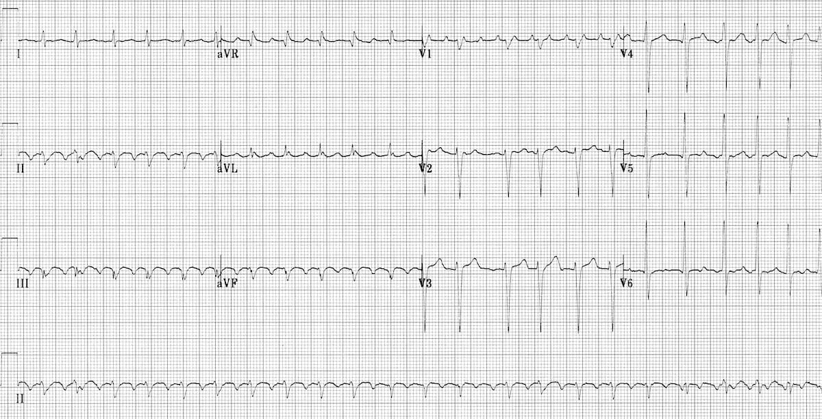
- Irregularly irregular rhythm
- No P waves
- Absence of an isoelectric baseline
- Variable ventricular rate
- QRS complexes usually <120 ms, unless there is pre-existing bundle branch block, an accessory pathway, or rate-related aberrant conduction
- Fibrillatory waves may be present — fine (amplitude <0.5 mm) or coarse (>0.5 mm) — and may mimic P waves, leading to misdiagnosis
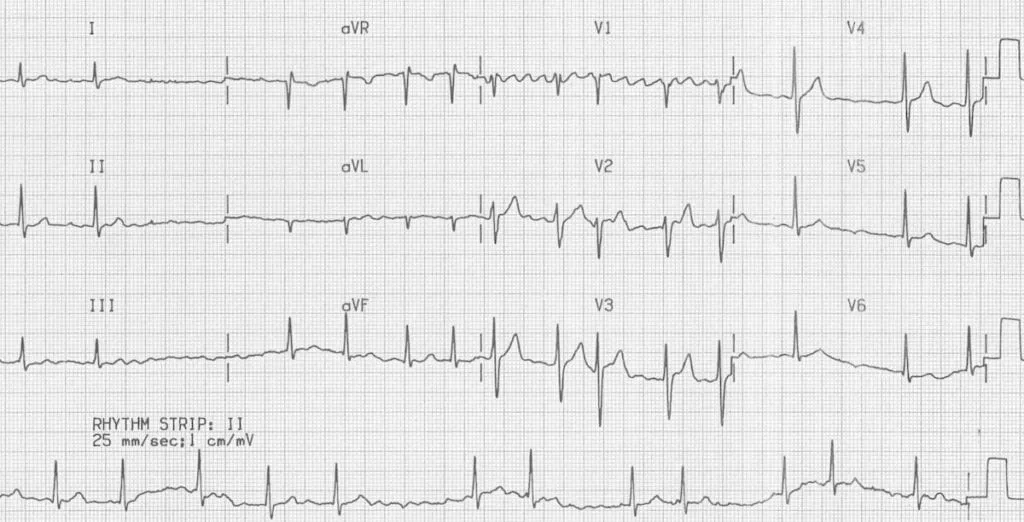
Atrial fibrillation: irregular ventricular response; coarse fibrillatory waves visible in V1.
Causes and risk factors
- Myocardial ischaemia (~15% of cases)
- Heart failure
- Elevated blood pressure
- Valvular heart disease
- Diabetes mellitus
- Obesity / excess body weight
- Obstructive sleep apnoea
- Hazardous alcohol use
- Hyperthyroidism
No cause is found in ~12% (“isolated” AF).
Investigations
- FBC, UEC, LFTs
- TSH — a thyroid-stimulating hormone test should be undertaken in all newly diagnosed AF (delay in acutely ill patients)
- Transthoracic echocardiogram — should be performed in all patients with newly diagnosed AF
- 12-lead ECG for all patients with AF
Screening and risk-factor management
The 2018 guideline added an explicit preventive/risk-factor dimension that the original notes didn’t cover:
- Opportunistic screening: opportunistic point-of-care screening in the clinic or community should be conducted in people aged 65 years or more.
- Weight: intensive weight management (target ≥10% body-weight loss, aiming for BMI <27) should be performed in overweight and obese individuals with AF.
- Sleep apnoea: screen for and manage OSA in recurrent symptomatic AF.
- Exercise: aerobic exercise is recommended in symptomatic AF to reduce AF burden.
- Identify and manage intercurrent comorbidities (hypertension, diabetes, heart failure, valvular disease, alcohol excess) as a core part of treatment.
Rate vs rhythm control — choosing a strategy
A rate-control or rhythm-control strategy should be selected, documented, communicated, and reviewed regularly for every AF patient. Rhythm control is particularly helpful in patients who remain symptomatic despite adequate rate control.
Stroke prevention
Risk stratification — CHA₂DS₂-VA
Thromboembolic risk is similar in persistent and paroxysmal AF, so anticoagulation should be considered for all these patients based on stroke risk, not AF pattern.
Subclinical (device-detected) AF — atrial high-rate episodes picked up incidentally on a pacemaker, ICD or implantable loop recorder, with no corresponding symptoms — is treated more cautiously than clinical AF. The 2024 ESC guideline is deliberately conservative here, reflecting two large trials (NOAH-AFNET 6 and ARTESiA) that found only a small anticoagulation benefit against an increased bleeding risk; routine anticoagulation is not recommended, and it is only considered case-by-case in patients with a high stroke risk and low bleeding risk.
Choose who to anticoagulate with the CHA₂DS₂-VA score — the sexless version of CHA₂DS₂-VASc is recommended for predicting stroke risk in AF:
| CHA₂DS₂-VA risk factor | Points |
|---|---|
| Congestive heart failure | +1 |
| Hypertension | +1 |
| Age ≥75 years | +2 |
| Diabetes mellitus | +1 |
| Previous Stroke, TIA or thromboembolism | +2 |
| Vascular disease (peripheral arterial disease, complex aortic plaque or prior MI) | +1 |
| Age 65–74 years | +1 |
- Score ≥2 → anticoagulate (oral anticoagulation is recommended)
- Score 1 → consider anticoagulation
- Score 0 → anticoagulation not recommended
Note the change from the older CHA₂DS₂-VASc: sex (female) is no longer scored as an independent point. Female sex only increases stroke risk when other risk factors are already present, so thresholds are now the same for everyone. The score should be re-evaluated yearly in low-risk patients who aren’t anticoagulated.
Antiplatelet therapy is not recommended for stroke prevention in N-VAF patients, regardless of stroke risk — aspirin is not an acceptable substitute for oral anticoagulation.
Bleeding risk
Assess bleeding risk (e.g. HAS-BLED) and, importantly, identify and correct reversible bleeding factors rather than using a high score to withhold anticoagulation outright.
Choice of anticoagulant
For non-valvular AF requiring anticoagulation, a DOAC (direct oral anticoagulant — apixaban, dabigatran or rivaroxaban; formerly “NOAC”) is recommended in preference to warfarin. DOACs have significantly lower rates of intracranial bleeding and haemorrhagic stroke than warfarin.
Apixaban (a factor Xa inhibitor):
- 5 mg BD
- 2.5 mg BD if at least 2 of: age ≥80, weight ≤60 kg, serum creatinine ≥133 µmol/L
- Avoid if calculated creatinine clearance <25 mL/min
DOAC contraindications / cautions:
- Severe renal impairment
- Clinically significant or active bleeding
- Hepatic disease with coagulopathy
- Interacting drugs (e.g. azole antifungals)
Warfarin — reserved for valvular AF (mechanical heart valve or moderate-to-severe mitral stenosis), where DOACs should not be used:
- Target INR 2–3 for non-valvular AF (if warfarin is used)
- Valvular AF — target may be higher (e.g. 2.5–3.5); seek specialist advice
Peri-procedural anticoagulation
- Minor bleeding-risk procedures (e.g. cataracts, minor dental work): DOAC often can be continued.
- Low bleeding-risk procedures: cease DOAC ~24 h prior (48 h if renal impairment).
- High bleeding-risk procedures: cease ~48 h prior (72–96 h if renal impairment).
- Bridging with LMWH/UFH is generally not needed for warfarin or DOAC patients at low-to-moderate stroke risk; bridging is reserved for very high risk (e.g. mechanical mitral valve).
Anticoagulation around cardioversion
Oral anticoagulation should be used for a minimum of 4 weeks following cardioversion (electrical or pharmacological), regardless of the pre-cardioversion strategy.
Rate control
Aim for a resting ventricular rate <110 bpm (lenient control) — documentation of adequacy of rate control (<110 bpm) should be performed at regular intervals in asymptomatic patients without heart failure.
First-line agents — beta-blockers, digoxin and non-dihydropyridine calcium-channel antagonists should be the first-line agents for long-term rate control:
- Beta-blocker — e.g. atenolol 25 mg daily (up to 100 mg), or metoprolol tartrate 25 mg BD (up to 100 mg BD)
- Non-dihydropyridine CCB — diltiazem MR 180 mg daily (up to 360 mg), or verapamil — but avoid in LV systolic dysfunction (EF <40%)
- In LV dysfunction (EF <40%), use a heart-failure beta-blocker instead: carvedilol, bisoprolol, nebivolol or metoprolol succinate
- Digoxin — second-line, or an add-on; useful adjunct where first-line agents are insufficient. Monitor serum concentration (goal <1.2 ng/mL)
- Amiodarone — useful for acute rate control in LV systolic dysfunction, or when other agents fail (200 mg daily; or 300 mg IV over 30–60 min acutely), but not a first-line agent for chronic rate control given its toxicity profile
If pharmacological rate control fails, AV-node ablation after permanent pacemaker implantation can be considered.
Rhythm control
Acute cardioversion
- Haemodynamically unstable AF → urgent electrical cardioversion.
- Stable AF <48 hours → low risk of thromboembolism; an initial rate- or rhythm-control strategy may be used. If rhythm control is chosen, anticoagulate at the time of cardioversion and continue longer-term depending on thromboembolic risk.
- Stable AF >48 hours (or unknown duration) → increased left-atrial thrombus risk. Do not perform acute cardioversion unless left-atrial thrombus has been excluded (TOE) or the patient has had therapeutic anticoagulation for the preceding 3 weeks. Otherwise rate control is the preferred initial approach; anticoagulate for ≥4 weeks after cardioversion.
Remember the possibility of early cardioversion in AF of clear onset <48 hours — stroke risk rises with duration, so the earlier the better.
Electrical cardioversion — synchronised DC cardioversion is highly effective and has a higher success rate than pharmacological cardioversion; biphasic defibrillators are more effective than monophasic.
Pharmacological cardioversion:
- Flecainide — preferred in patients with normal LV function and no coronary disease (1.5–2 mg/kg, up to 150 mg, IV over 10 min). Give with an AV-nodal blocker to avoid 1:1 conduction if it organises to flutter.
- Amiodarone — preferred in LV systolic dysfunction (EF <40%) or coronary disease (300 mg IV over 1–2 h, then 900 mg IV over 24 h).
Long-term rhythm control
Difficult to maintain, but worthwhile if the patient is symptomatic.
- If normal LV function and no coronary disease — flecainide 50 mg BD, up to 150 mg BD (with a rate-limiting AV-nodal blocker such as diltiazem or a beta-blocker to reduce the risk of 1:1 atrial-flutter conduction)
- Sotalol — 40 mg up to 160 mg BD; watch the QT interval and stop if QTc >500 ms
- Amiodarone — 200 mg TDS for 1 week, then 200 mg BD for 1 week, then 200 mg daily; second-line generally, but first-line where there is LV systolic dysfunction, moderate LVH or coronary disease
Catheter ablation
Increasingly used, and moving earlier in the pathway rather than being a last resort:
- Catheter ablation should be considered for symptomatic paroxysmal or persistent AF refractory or intolerant to at least one Class I or III antiarrhythmic medication.
- It can also be considered before antiarrhythmic therapy in selected symptomatic patients, and in selected patients with heart failure with reduced ejection fraction.
- The 2024 ESC AF guideline gives ablation a still more prominent first-line role in selected patients, under its AF-CARE framework — Comorbidity and risk-factor management, Avoid stroke and thromboembolism, Rhythm and rate control, Evaluation and dynamic reassessment (see the update block below).
Atrial flutter
- The ECG shows a regular saw-tooth baseline with a ventricular rate around 150 and narrow QRS complexes (2:1 AV block); it is often misdiagnosed as SVT.
- Greater AV block gives a ventricular rate of ~100 (3:1) or ~75 (4:1). Rarely, 1:1 conduction gives a rate of ~300.
- Commonly reverts with a low-energy DC shock or overdrive pacing, and is often relatively insensitive to antiarrhythmic drugs.
- Electrical cardioversion is often performed earlier than in AF.
- Options for rhythm control in atrial flutter include electrical cardioversion, cavotricuspid isthmus (CTI) ablation for typical flutter — CTI ablation is markedly more effective than antiarrhythmic drugs for preventing recurrence.
- Stroke-prevention (anticoagulation) principles are the same as for AF, and otherwise the management of atrial flutter and AF is generally the same.